Hip Biomechanics
The hip functions to support the weight of the upper body and transmit forces to the lower extremities. The ball and socket configuration allows for stability and mobility.
Structure
- See also: Hip Joint
Acetabulum
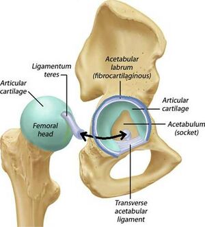
The acetabulum is not completely spherical due to the acetabular notch in its inferior region, which makes it fundamentally horseshoe shaped. Articular cartilage, covering the surface of the acetabulum, thickens peripherally and laterally, though most notably in the superior-anterior region of the dome.
The acetabular labrum, a fibrocartilaginous lip encircling and deepening the acetabulum, blends with the transverse acetabular ligament, which spans the acetabular notch and prevents inferior dislocation of the femoral head. The cavity of the acetabulum faces obliquely forward, outward, and downward. The average value is approximately 20°; pathologic increases in the angle of acetabular anteversion are associated with decreased joint stability and an increased likelihood of anterior dislocation of the head of the femur (see Hip Joint).
Unlike capsular tissue, labral tissue is made up predominantly of fibrocartilage. Arthroscopic visualization of damaged labral tissue has shown more extensive penetration of vascular tissue throughout the entire labral structure, which suggests more healing potential than previously believed. The labrum plays a role in containing the femoral head in extremes of motion, particularly in flexion.
It is believed that there is interaction between the intra-articular fluid and the labrum, which decreases peak pressures in the joint. The labrum also plays a role in maintaining a vacuum within the joint space, which is produced by the acetabular fossa, a depression in the centre of the acetabulum.
Femoral Head
The femoral head, the convex component of the ball-and-socket configuration of the hip joint, forms two-thirds of a sphere. The articular cartilage covering the femoral head is thickest on the medial-central surface surrounding the fovea into which the ligamentum teres attaches and thinnest toward the periphery. The load-bearing area is concentrated at the periphery of the lunate surface of the femoral head at smaller loads, but it shifts to the center of the lunate surface and the anterior and posterior horns as loads increase.
Femoral Neck
The inclination angle is about 140° to 150° at birth and gradually reduces to approximately 125°, with a range of 45° (90°–135°) in adulthood. An angle greater than 125° produces a condition known as coxa valga, while an angle less than 125° is known as coxa vara.
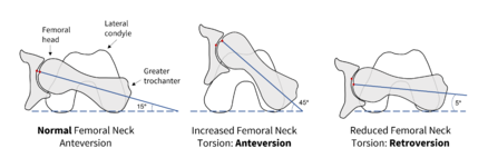
The torsion angle reflects medial rotary migration of the lower limb bud that occurs in fetal development; it is commonly estimated at 40° in newborns, but decreases substantially in the first two years of life. A torsion angle of between 10° and 20° is considered normal. Angles greater than 12°, known as anteversion, cause a portion of the femoral head to be uncovered and create a tendency toward internal rotation of the leg during gait to keep the femoral head in the acetabular cavity. Retroversion, an angle of less than 12°, produces a tendency toward external rotation of the leg during gait. Both are fairly common during childhood and are usually outgrown.
The interior of the femoral head and neck are composed of cancellous bone with trabeculae organized into medial and lateral trabecular systems (See Image). The forces and stresses on the femoral head—most specifically the joint reaction force—parallel the trabeculae of the medial system.
The hip capsule, composed of three capsular ligaments, is an important stabilizer of the hip joint, especially in extremes of motion, where it acts as a check rein to prevent dislocation (Johnston et al., 2007). The capsular ligament is made up of three reinforcing ligaments—two found anteriorly and one posteriorly. The capsule is thickened anterosuperiorly, where the predominant stresses occur, and is relatively thin and loosely attached posteroinferiorly. Due to the rotation that occurs in fetal development of the hip joint, the capsular ligaments are coiled around the femoral neck in a clockwise direction, meaning that they are tightest in combined extension and medial rotation of the hip joint, which further coils the ligaments, and loosest in flexion and lateral rotation.
Kinematics
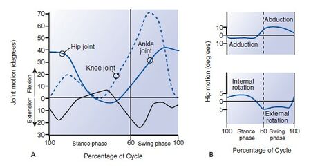
Hip motion takes place in all three planes: sagittal (flexion-extension), frontal (abduction-adduction), and transverse (internal-external rotation). Motion is greatest in the sagittal plane, where the range of flexion is from 0° to approximately 140° and the range of extension is from 0° to 15°. The range of abduction is from 0° to 30°, whereas that of adduction is somewhat less, from 0° to 25°. External rotation ranges from 0° to 90° degrees and internal rotation from 0° to 70° when the hip joint is flexed. Less rotation occurs when the hip joint is extended because of the restricting function of the soft tissues.
Maximal motion in the sagittal plane (hip flexion) was needed for tying the shoe and bending down to squat to pick up an object from the floor. The greatest motion in the frontal and transverse planes was recorded during squatting and during shoe tying with the foot across the opposite thigh. The values obtained for these common activities indicate that hip flexion of at least 120° and external rotation of at least 20° are necessary for carrying out daily activities in a normal manner.
- Stance phase- 60% , hip abducted, externally rotated
- Swing phase- 40%, hip adducted, internally rotated.
Easier to calculate forces when two leg stance, as don’t have to include abductor muscle forces, just ground reactive forces. 1 leg stance--- more complicated, need to include other forces.
Single leg stance is about 3 times body weight.
In men, two peak forces were produced during the stance phase when the abductor muscles contracted to stabilize the pelvis. One peak of approximately four times body weight occurred just after heel-strike, and a large peak of approximately seven times body weight was reached just before toe-off. In women, the force pattern was the same but the magnitude was somewhat lower, reaching a maximum of only approximately four times body weight at late stance phase. several factors: a wider female pelvis, a difference in the inclination of the femoral neck-to-shaft angle, a difference in footwear, and differences in the general pattern of gait. It was found that females walked with greater adduction angles at the hip, which contributed to the greater adduction moment, suggesting a narrower step width relative to pelvic width. This implies that the hip joint stress for the female population is higher, not only in static situations, but also in dynamic activities, as compared with males.
Neumann found that use of a cane on the contralateral side of the affected hip joint, with careful instructions to use with near maximal effort, could reduce the muscle activity by 42%. This calculates to a reduction of approximately one times body weight: from 2.2 times body weight with a cane to 3.4 times body weight without.
The magnitude of the hip joint reaction force is influenced by the ratio of the abductor muscle force lever arm to the gravitational force lever arm. A low ratio (closer to zero) yields a greater joint reaction force than does a high ratio (one closer to 0.8). During a gait cycle, the hip joint reaction force experienced in stance phase is equal to or greater than three to six times body weight, while being approximately equal to body weight during swing phase.
See Also
References
Nordin, Margareta, and Victor H. Frankel. Basic biomechanics of the musculoskeletal system. Philadelphia: Wolters Kluwer Health/Lippincott Williams & Wilkins, 2012.


