Neuronopathies: Difference between revisions
mNo edit summary |
|||
| Line 162: | Line 162: | ||
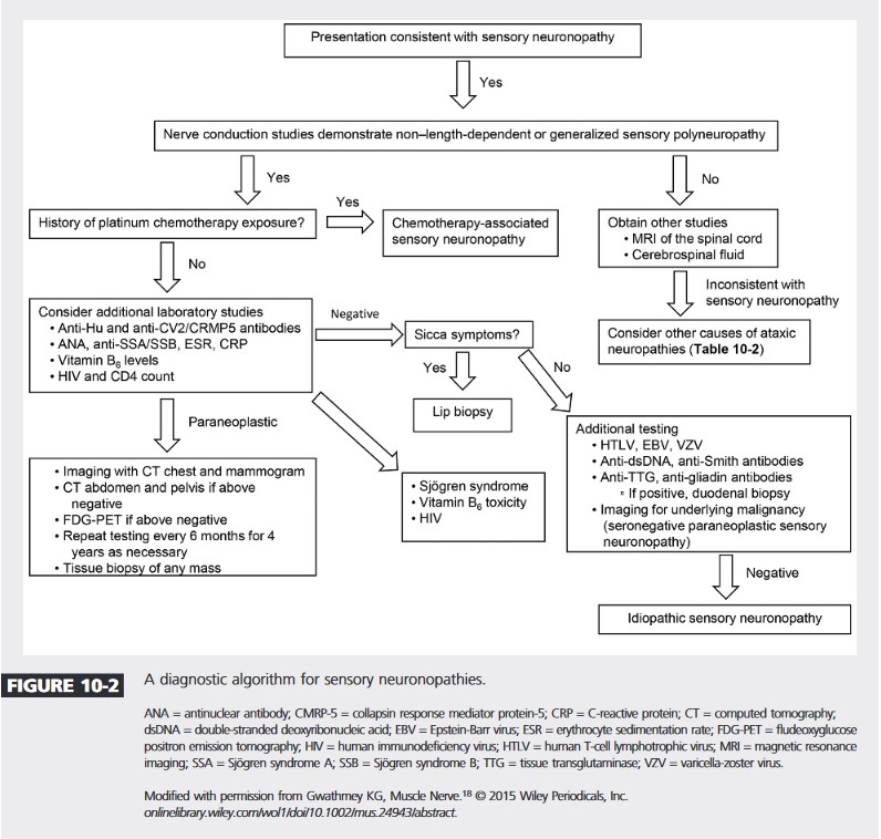
MRI findings of T2 hyperintensity of the dorsal column is strongly supportive of the diagnosis. The dorsal column contains the central afferent projections of the DRG. This finding is not present in the majority of patients.<ref name=":1" />[[File:Sensory neuronopathy algorithm continuum.jpg|center|frame|Diagnostic algorithm from 2016 Continuum article<ref>{{Cite journal|last=Gwathmey|first=Kelly Graham|date=2017-10|title=Sensory Polyneuropathies|url=https://journals.lww.com/00132979-201710000-00016|journal=CONTINUUM: Lifelong Learning in Neurology|language=en|volume=23|issue=5|pages=1411–1436|doi=10.1212/CON.0000000000000518|issn=1538-6899}}</ref>]] | MRI findings of T2 hyperintensity of the dorsal column is strongly supportive of the diagnosis. The dorsal column contains the central afferent projections of the DRG. This finding is not present in the majority of patients.<ref name=":1" />[[File:Sensory neuronopathy algorithm continuum.jpg|center|frame|Diagnostic algorithm from 2016 Continuum article<ref>{{Cite journal|last=Gwathmey|first=Kelly Graham|date=2017-10|title=Sensory Polyneuropathies|url=https://journals.lww.com/00132979-201710000-00016|journal=CONTINUUM: Lifelong Learning in Neurology|language=en|volume=23|issue=5|pages=1411–1436|doi=10.1212/CON.0000000000000518|issn=1538-6899}}</ref>]] | ||
== Resources == | |||
{{Members link}} | |||
==References== | ==References== | ||
Revision as of 20:39, 27 November 2023
Neuronopathy means the cell body is affected rather than the myelin or axon as in peripheral neuropathy. This article is focused on the sensory neuronopathies where there is primary degeneration of the dorsal root ganglion and trigeminal ganglion sensory neurons. It is a rare type of polyneuropathy.
Classification
The classification of neuropathic disorders is as follows
- Neuronopathies (pure sensory or pure motor or autonomic)
- Sensory neuronopathies (ganglionopathies)
- Motor neuronopathies (motor neuron disease)
- Autonomic neuropathies
- Peripheral neuropathies (usually sensorimotor)
- Myelinopathies
- Axonopathies
- Large- and small-fibre
- Small-fibre
Anatomy
The dorsal root ganglion (DRG) contains the cell bodies of sensory pseudo-unipolar neurons. The dorsal roots contain their efferent projections. Capillaries supply the DRG and in doing so cause fenestration and thereby loosening the blood-nerve barrier. This increases susceptibility to antibodies and toxins.
There are two populations of neurons in the DRG. These are the large-light cells with their Aβ and Aδ fibre projections. The other population, which are the majority of the cells, are the small-dark cells and their unmyelinated C fibres. The afferent projections of the larger neurons containing proprioceptive and tactile sensation traverse the posterior column.
Epidemiology
It is more common in women. The median age of onset is 54. Common comorbidities include diabetes, excessive alcohol intake, smoker, malignancy, autoimmune disease.[1]
Clinical Features
The sensory neuronopathies classically initially (but not always initially[1]) present with early onset sensory ataxia, potentially secondary to proximal muscle spindle and joint denervation.[2] This reflects damage to the large-fibre neurons. Note, early onset sensory ataxia is not unique to sensory ganglionopathy. It can also be caused by demyelinating large fibre neuropathy and pathology localised to the dorsal column. With severe proprioceptive loss the patient may have "pseudoathetosis" of the fingers and toes. If sensory ataxia doesn't present initially, it almost always eventually manifests at full development. The sensory ataxia is the main cause of disability.
If the small and medium sized nerves are also involved then the patient may experience neuropathic pain (positive sensory symptoms). Positive features may include hyperaesthesia and allodynia.[2]
This leads to the other classic feature being that the negative symptoms (numbness) and positive symptoms often present with a non-length-dependent and multifocal pattern. This differentiates them from typical length dependent axonal sensory polyneuropathies. Misdiagnosis can occur if the examiner is testing sensation from distal to proximal and stops at the presumed sensory level, thereby missing the multifocal presentation. With severe diffuse involvement of DRGs the clinical pattern may mimic a length-dependent distribution.[1]
Symptoms can initially affect the upper limbs, lower limbs, or all four limbs. Symptoms can be asymmetrical or symmetrical. [1]
Vestibular dysfunction can occur, such as in CANVAS syndrome. CANVAS patients are also more likely to have chronic neurogenic cough, which can help distinguish this group.
Power is usually normal, but patients may appear weak due to lack of proprioceptive feedback and loss of sensory input from denervated spindles and Golgi tendon organs.
The muscle stretch reflexes are often unelicitable.
| Symptoms in % (n=53) | At onset | At full development | At any time |
|---|---|---|---|
| Pain | 17 (32) | 32 (59) | |
| Hypoaesthesia | 36 (68) | 53 (99) | |
| Paraesthesia | 27 (51) | 24 (44) | |
| Ataxia | 17 (32) | 51 (94) | |
| Pseudoaethosis | 0 (0) | 12 (22) | |
| Dry eyes and mouth | 4 (7) | 11 (20) | |
| Autonomic symptoms | 10 (19) | 14/52 (27) | |
| Orthostatic hypotension | 4 (7) | 6 (11) | |
| Gastrointestinal dysfunction | 4 (7) | 8 (15) | |
| Dysphagia | 2 (4) | 5 (9) | |
| Erectile dysfunction | 1 (2) | 2 (4) | |
| Bladder dysfunction | 0 (0) | 5 (9) | |
| Sweating dysfunction | 1 (2) | 4 (7) | |
| Pupil abnormalities | 1 (2) | 3/53 (6) | |
| Vestibular dysfunction | 5 (9) | ||
| Cough | 6/53 (11) | ||
| Facial involvement | 11 (20) | 15 (28) | |
| Motor involvement | 15 (28) | ||
| Eye movement abnormalities | 8 (15) | ||
| Generalised areflexia | 28/52 (54) |
Aetiology
- Immune mediated disease: Sjogren syndrome, systemic lupus erythematosus, autoimmune hepatitis, celiac disease
- Paraneoplastic disease: Small-cell lung cancer, bronchial carcinoma, breast cancer, ovarian cancer, Hodgkin lymphoma, transitional cell bladder cancer, prostate cancer, malignant mixed M€ullerian tumor, neuroendocrine tumor, sarcoma
- Viral infections: HIV, EBV, VZV, HTLV-1
- Vitamin B6
- Intoxication
- Neurotoxic drugs: pyridoxine, cisplatin, carboplatin, oxaliplatin
- Hereditary and degenerative disorders: hereditary sensory autonomic neuropathy (HSAN), Charcot-Marie-tooth disease type 2B (CMT2B), sensory ataxia neuropathy with dysarthria and ophthalmoparesis (SANDO), facial-onset sensory motor neuronopathy (FOSMN), and Cerebellar ataxia, neuropathy, vestibular areflexia syndrome (CANVAS)[2]
- Idiopathic (~50%)[1]
Investigations
The first step is usually nerve conduction studies. This is to identify severely reduced or absent SNAPs indicating severe, generalised, non-length-dependent sensory neuropathy. Upper extremity sensory nerves may be affected to a greater degree than lower extremity, unlikely axonal neuropathies.
MRI findings of T2 hyperintensity of the dorsal column is strongly supportive of the diagnosis. The dorsal column contains the central afferent projections of the DRG. This finding is not present in the majority of patients.[1]

Resources
References
- ↑ 1.0 1.1 1.2 1.3 1.4 1.5 1.6 Sancho Saldaña, Agustin; Mahdi‐Rogers, Mohamed; Hadden, Robert David (2021-03). "Sensory neuronopathies: A case series and literature review". Journal of the Peripheral Nervous System (in English). 26 (1): 66–74. doi:10.1111/jns.12433. ISSN 1085-9489. Check date values in:
|date=(help) - ↑ 2.0 2.1 2.2 Gwathmey, Kelly Graham (2016-01). "Sensory neuronopathies". Muscle & Nerve. 53 (1): 8–19. doi:10.1002/mus.24943. ISSN 1097-4598. PMID 26467754. Check date values in:
|date=(help) - ↑ Gwathmey, Kelly Graham (2017-10). "Sensory Polyneuropathies". CONTINUUM: Lifelong Learning in Neurology (in English). 23 (5): 1411–1436. doi:10.1212/CON.0000000000000518. ISSN 1538-6899. Check date values in:
|date=(help)

