File list
From WikiMSK
This special page shows all uploaded files.
| Date | Name | Thumbnail | Size | User | Description | Versions |
|---|---|---|---|---|---|---|
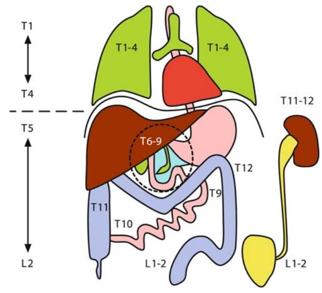
| 10:57, 21 August 2021 | Visceral referred pain.jpg (file) |  |
42 KB | Jeremy | The topography of the thoracic and abdominal viscera. The viscera above the diaphragm are innervated by T1-4, while the viscera below the diaphragm are innervated by T5-L2. Copyright Borowczyk J. (2007) Visceral Referred Pain. In: Schmidt R., Willis W. (eds) Encyclopedia of Pain. Springer, Berlin, Heidelberg. https://doi.org/10.1007/978-3-540-29805-2_4801 | 1 |
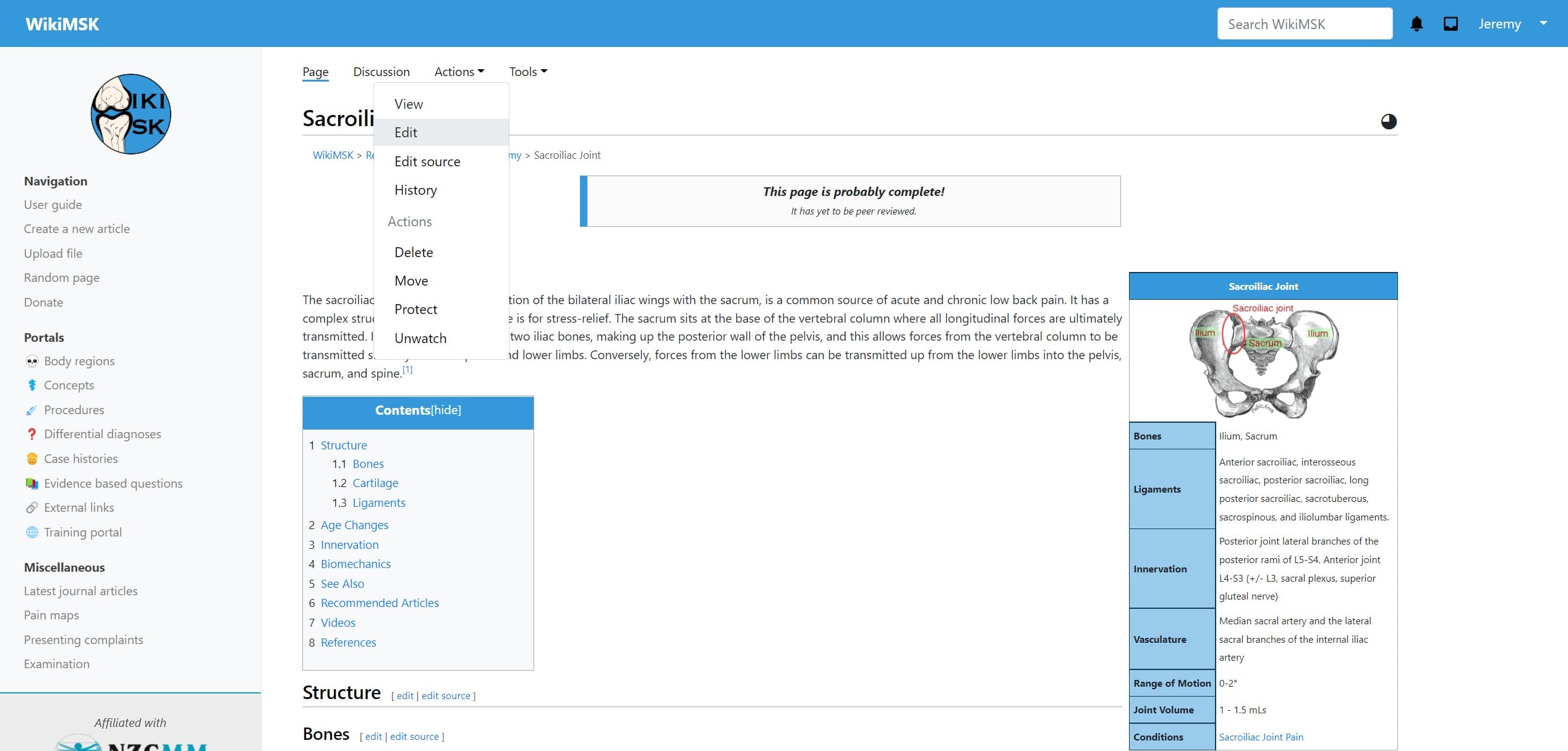
| 14:15, 20 March 2022 | Visual editor location.jpg (file) |  |
177 KB | Jeremy | 1 | |
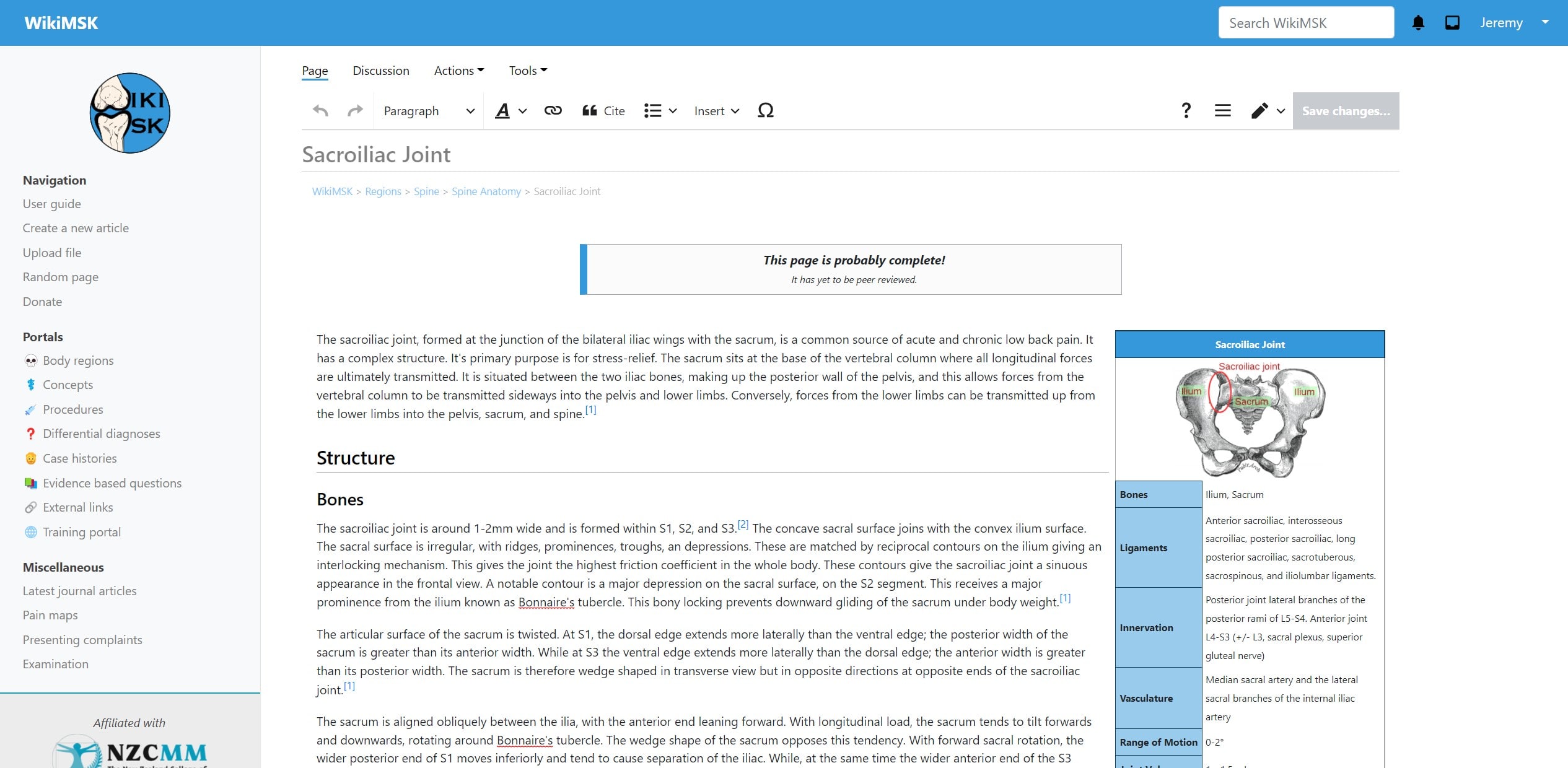
| 14:47, 20 March 2022 | Visual editor view.jpg (file) |  |
317 KB | Jeremy | 1 | |
| 21:15, 26 September 2020 | Volar-wrist.jpg (file) |  |
42 KB | Jeremy | File uploaded with MsUpload | 1 |
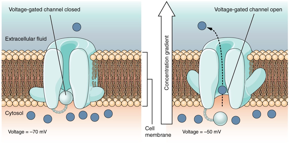
| 18:56, 12 July 2021 | Voltage-gated-channels.jpg (file) |  |
131 KB | Jeremy | File uploaded with MsUpload | 1 |
| 18:02, 19 May 2023 | Voltage gated sodium channels and blockers - Li 2019.pdf (file) | 2.07 MB | Jeremy | File uploaded with MsUpload | 1 | |
| 08:45, 8 April 2021 | WIKIMSK hero.png (file) | 181 KB | Jeremy | File uploaded with MsUpload | 1 | |
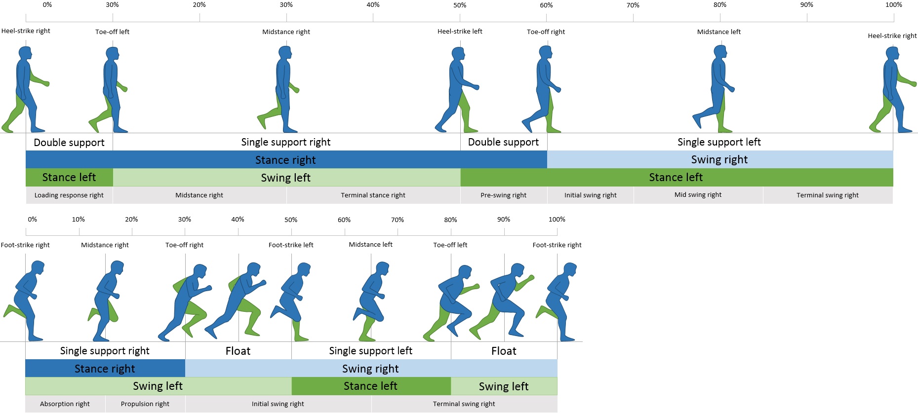
| 15:06, 13 March 2022 | Walk and run cycle.jpg (file) |  |
233 KB | Jeremy | From https://commons.wikimedia.org/wiki/File:Walk_and_run_cycle.jpg | 1 |
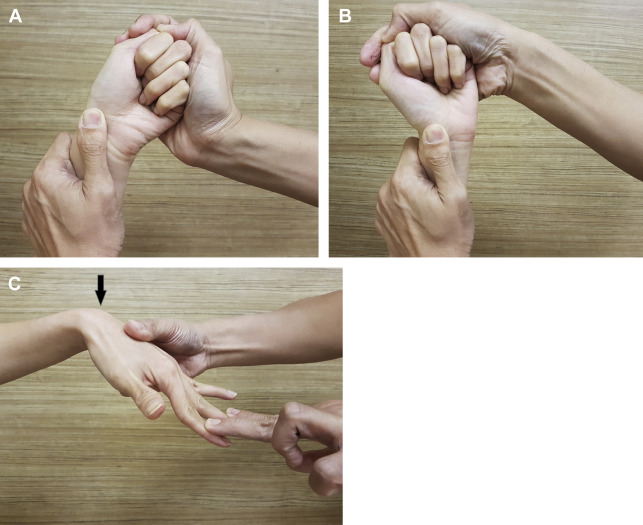
| 14:38, 16 September 2021 | Watson and finger extension tests.jpg (file) |  |
97 KB | Jeremy | A and B: watson tests C:finger extension test Tan DMK, Lim JX. Treatment of Carpal Instability and Distal Radioulnar Joint Instability. Clin Plast Surg. 2019 Jul;46(3):451-468. doi: 10.1016/j.cps.2019.03.006. PMID: 31103089. | 1 |
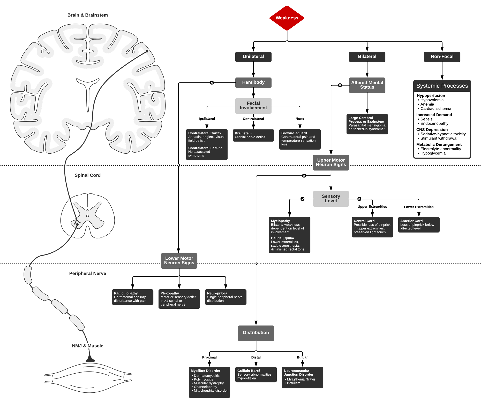
| 07:42, 7 May 2022 | Weakness algorithm.png (file) |  |
133 KB | Jeremy | From https://ddxof.com/weakness-2/ | 1 |
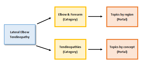
| 21:19, 28 June 2020 | Website Structure.PNG (file) |  |
11 KB | Jeremy | File uploaded with MsUpload | 1 |
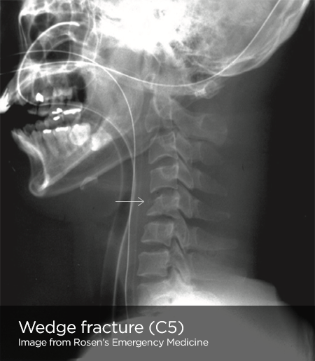
| 06:51, 7 May 2022 | Wedge.png (file) |  |
136 KB | Jeremy | Radiopaedia | 1 |
| 07:55, 8 April 2021 | Welcome to wikimsk.png (file) |  |
301 KB | Jeremy | File uploaded with MsUpload | 1 |
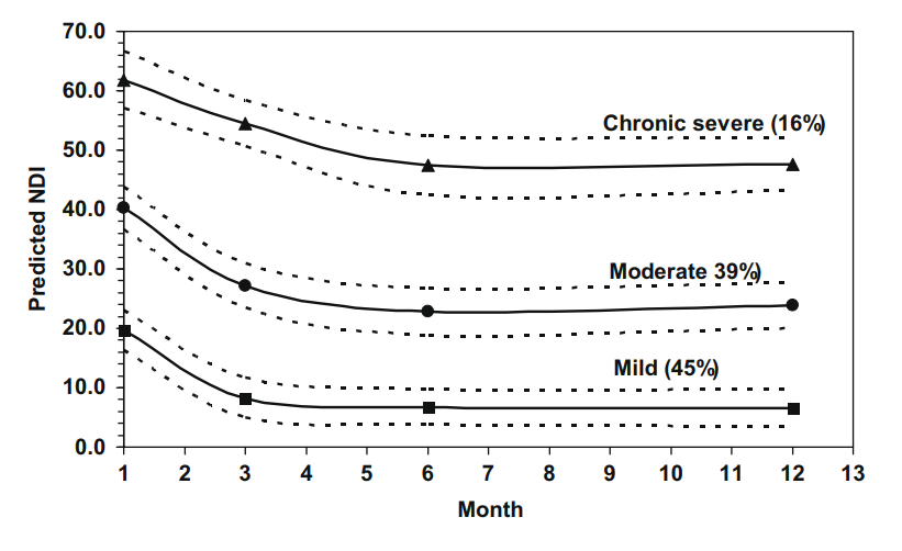
| 13:46, 5 July 2021 | Whiplash NDI trajectories.png (file) |  |
60 KB | Jeremy | File uploaded with MsUpload | 1 |
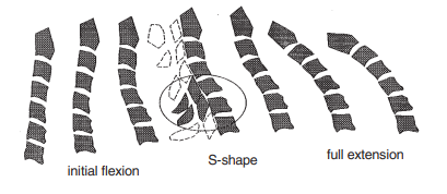
| 17:17, 27 June 2021 | Whiplash cervical spine S shaped.png (file) |  |
44 KB | Jeremy | File uploaded with MsUpload | 1 |
| 17:45, 23 May 2021 | Whiplash cervical spine motion.png (file) |  |
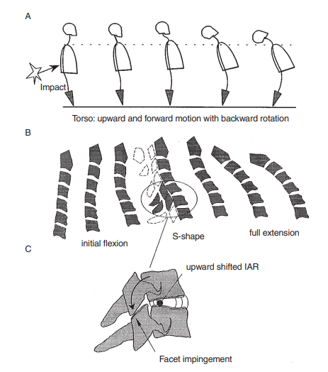
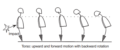
99 KB | Jeremy | A: The torso motion during simulated rear-end impact. The subject’s torso pushes upward and forward from the seat back with backward rotation. This torso motion and head inertia produces the force to the neck. B: in situ whiplash motion. The backward rotation starts from C7 and gradually transfers to the upper vertebrae. The cervical spine moves in a whiplash-like manner. During this process, the cervical spine shows flexion in the early phase and is S-shaped in the middle phase. C: The hyp... | 1 |
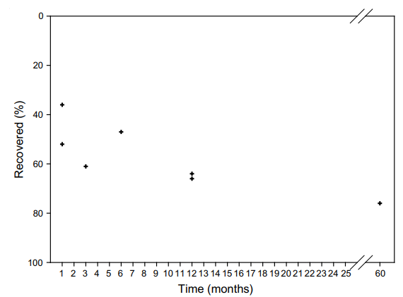
| 08:48, 5 July 2021 | Whiplash recovery rate.png (file) |  |
19 KB | Jeremy | File uploaded with MsUpload | 1 |
| 17:17, 27 June 2021 | Whiplash torso movement.png (file) |  |
22 KB | Jeremy | File uploaded with MsUpload | 1 |
| 09:19, 31 August 2020 | Why I Pursue Discogenic Pain - Bogduk.pdf (file) | 1.97 MB | Jeremy | File uploaded with MsUpload | 1 | |
| 09:12, 31 August 2020 | Why do an MSK Exam.pptx (file) | 788 KB | Jeremy | File uploaded with MsUpload | 1 | |
| 16:02, 19 August 2021 | Why most research findings are false - ioannidis 2005.pdf (file) | 250 KB | Jeremy | File uploaded with MsUpload Category:Statistics | 1 | |
| 05:42, 31 March 2022 | WikiMSK2894-fracture.png (file) |  |
6 KB | Jeremy | Uploaded with SimpleBatchUpload | 1 |
| 17:33, 31 August 2020 | WikiMSK Presentation.pptx (file) | 2.91 MB | Jeremy | File uploaded with MsUpload | 1 | |
| 17:59, 20 March 2022 | WikiMSK logo.png (file) | 48 KB | Jeremy | smaller | 2 | |
| 20:21, 15 March 2022 | Wiki email.png (file) | 9 KB | Jeremy | 2 | ||
| 20:28, 15 March 2022 | Wiki email info.png (file) | 9 KB | Jeremy | 2 | ||
| 17:54, 11 August 2020 | Williams2019 - intrapartum lesions lumbar potion of lumbosacral plexus.pdf (file) | 778 KB | Jeremy | File uploaded with MsUpload | 1 | |
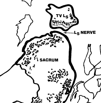
| 17:50, 25 April 2021 | Wiltse far out syndrome.png (file) |  |
34 KB | Jeremy | File uploaded with MsUpload | 1 |
| 14:35, 3 April 2022 | Woman.png (file) |  |
19 KB | Jeremy | 1 | |
| 18:06, 27 July 2020 | Woolf2011 - Central sensitisation.pdf (file) | 485 KB | Jeremy | File uploaded with MsUpload | 1 | |
| 18:06, 27 July 2020 | Woolf2014 - Nociceptive amplification naming.pdf (file) | 125 KB | Jeremy | File uploaded with MsUpload | 1 | |
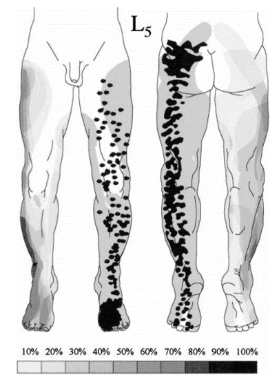
| 11:21, 8 August 2021 | Woolf L5 dermatome.png (file) |  |
77 KB | Jeremy | File uploaded with MsUpload | 1 |
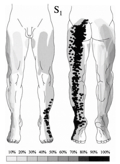
| 11:21, 8 August 2021 | Woolf S1 dermatome.png (file) |  |
73 KB | Jeremy | File uploaded with MsUpload | 1 |
| 19:24, 20 October 2022 | Word.png (file) |  |
2 KB | Jeremy | 1 | |
| 08:48, 13 September 2020 | Wray2020 the art of the deal.pdf (file) | 101 KB | Jeremy | File uploaded with MsUpload | 1 | |
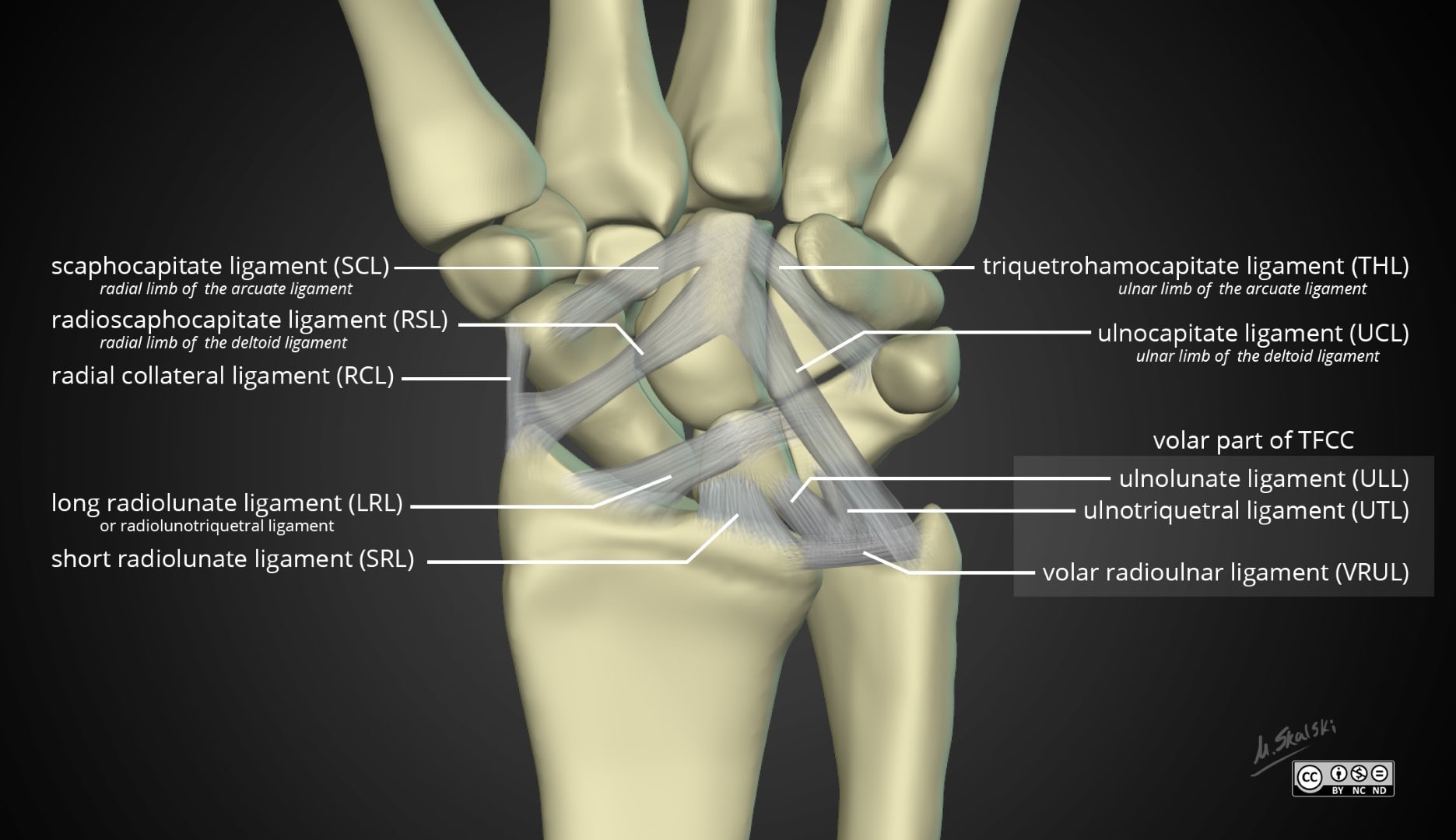
| 07:18, 6 February 2022 | Wrist-anatomy-extrinsic-ligaments.jpg (file) |  |
150 KB | Jeremy | Case courtesy of Dr Matt Skalski, <a href="https://radiopaedia.org/?lang=gb">Radiopaedia.org</a>. From the case <a href="https://radiopaedia.org/cases/43845?lang=gb">rID: 43845</a> | 1 |
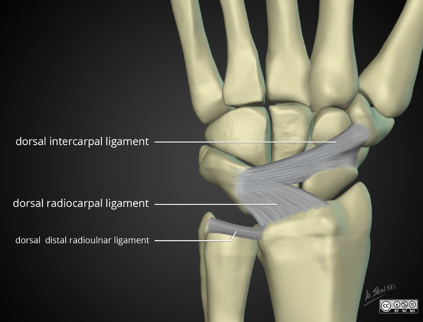
| 07:22, 6 February 2022 | Wrist-anatomy-extrinsic-ligaments dorsal.jpg (file) |  |
90 KB | Jeremy | Case courtesy of Dr Matt Skalski, <a href="https://radiopaedia.org/?lang=gb">Radiopaedia.org</a>. From the case <a href="https://radiopaedia.org/cases/43845?lang=gb">rID: 43845</a> | 1 |
| 21:53, 2 March 2022 | Wrist.png (file) |  |
4 KB | Jeremy | 1 | |
| 18:30, 5 May 2022 | Wrist Arthrocentesis.jpg (file) |  |
56 KB | Jeremy | From https://wikem.org/wiki/File:Wrist_Arthrocentesis.jpg | 1 |
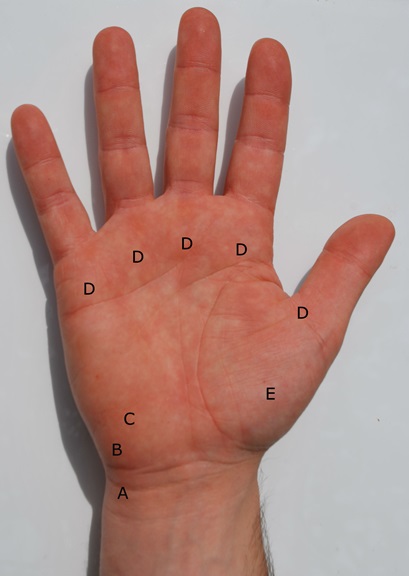
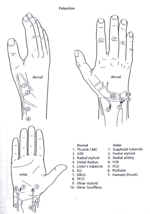
| 19:41, 30 June 2021 | Wrist palpation landmarks.png (file) |  |
68 KB | Jeremy | 2 | |
| 17:56, 9 September 2020 | Write.png (file) |  |
7 KB | Jeremy | File uploaded with MsUpload | 1 |
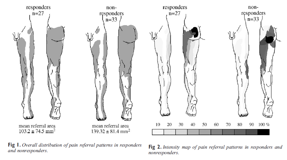
| 17:57, 16 April 2021 | Wurff SIJ maps.png (file) |  |
176 KB | Jeremy | File uploaded with MsUpload | 1 |
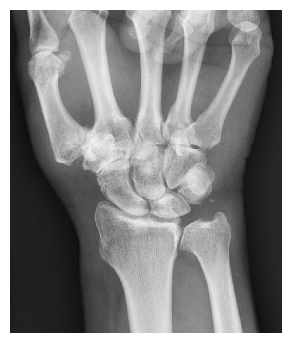
| 16:41, 14 June 2021 | Xray STT joint osteoarthritis.jpg (file) |  |
72 KB | Jeremy | An example of STT joint arthritis stage 3 according to the classification system used. There is also thumb trapeziometacarpal arthritis and distal radioulnar joint arthritis. There is no evidence of radiocarpal arthritis. {{#pmid:22957252}} | 1 |
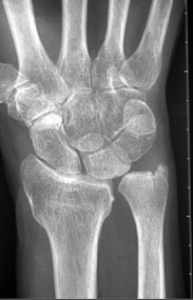
| 18:54, 3 April 2022 | Xray positive ulnar variance.png (file) |  |
62 KB | Jeremy | X-ray showing positive ulnar variance. (from radiopaedia) | 1 |
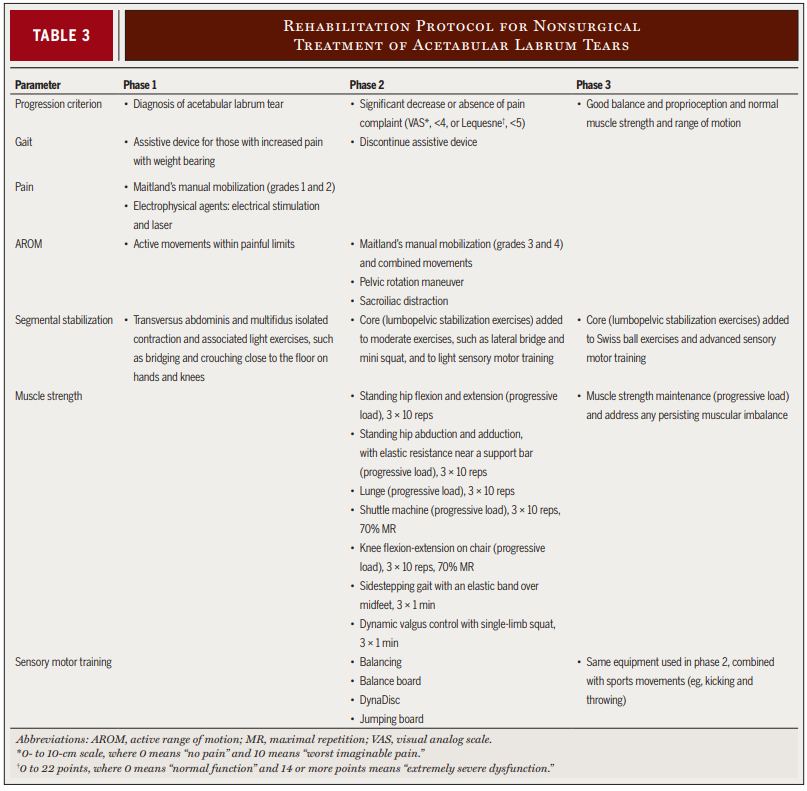
| 19:49, 14 July 2020 | Yazbek progression program.PNG (file) |  |
147 KB | Jeremy | File uploaded with MsUpload | 1 |
| 18:06, 27 July 2020 | Yunus2008 - Central sensitivity syndrome.pdf (file) | 337 KB | Jeremy | File uploaded with MsUpload | 1 | |
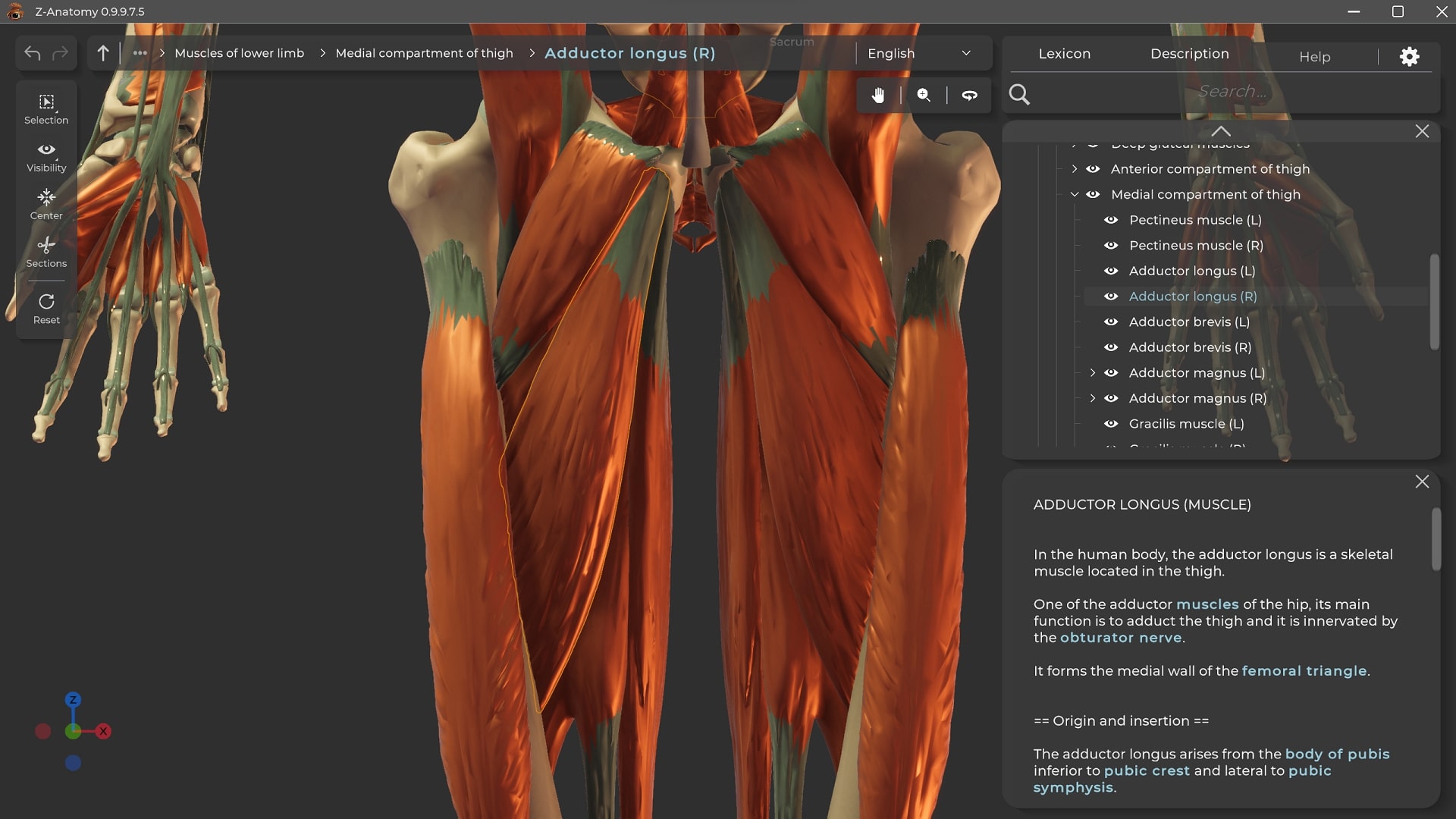
| 21:11, 6 November 2022 | Z anatomy screenshot.jpg (file) |  |
231 KB | Jeremy | 1 | |
| 14:47, 13 September 2020 | Zejnullahu2019 off trail on track an exercise in clinical reasoning.pdf (file) | 511 KB | Jeremy | File uploaded with MsUpload | 1 | |
| 16:47, 14 June 2022 | Zip.png (file) |  |
3 KB | Jeremy | 1 | |
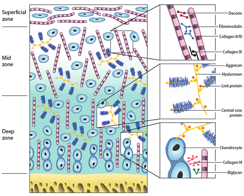
| 12:36, 31 July 2021 | Zones and constituents of articular cartilage.png (file) |  |
196 KB | Jeremy | Zones and constituents of articular cartilage. A schematic representation of the different matrix components and their organization throughout articular cartilage. Baer K, Kieser S, Schon B, Rajendran K, Ten Harkel T, Ramyar M, Löbker C, Bateman C, Butler A, Raja A, Hooper G, Anderson N, Woodfield T. Spectral CT imaging of human osteoarthritic cartilage via quantitative assessment of glycosaminoglycan content using multiple contrast agents. APL Bioeng. 2021 Apr 1;5(2):026101. doi: 10.1063... | 1 |

