Transverse Tarsal Joint (Chopart's Joint): Difference between revisions
No edit summary |
mNo edit summary |
||
| Line 19: | Line 19: | ||
ATFL injuries can occur along with injury to the calcaneocuboid ligament. Pain may be reproduced by fixing the rearfoot in dorsiflexion and eversion, and then inverting and adducting the forefoot. | ATFL injuries can occur along with injury to the calcaneocuboid ligament. Pain may be reproduced by fixing the rearfoot in dorsiflexion and eversion, and then inverting and adducting the forefoot. | ||
==Notes== | |||
<references group="notes"/> | <references group="notes"/> | ||
[[Category:Foot and Ankle Anatomy]] | [[Category:Foot and Ankle Anatomy]] | ||
Revision as of 18:51, 17 July 2021
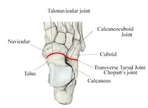
The transverse tarsal joint or midtarsal joint or Chopart's joint is formed by the articulation of the calcaneus with the cuboid (the calcaneocuboid joint), and the articulation of the talus with the navicular (the talocalcaneonavicular joint). This pair of joints separates the midfoot from the rearfoot (i.e. calcaneus and talus) by allowing the midfoot to move independently of the rearfoot.
Movement
The joint has the ability to perform the most pure form of pronation[notes 1] and supination[notes 2]. It allows the midfoot to conform to a variety of different positions depending on the terrain. The joint is considered to be part of the same functional unit as the subtalar joint because they share a common axis of rotation with contribution to inversion and eversion of the foot.
Ligaments
The calcaneocuboid joint, which is part of the lateral longitudinal arch of the foot and takes the whole bodyweight, is stabilised by the below ligaments and is also reinforced by the peroneus longus tendon. This joint is part of the lateral longitudinal arch of the foot.
- Plantar calcaneocuboid ligament. This passes from the anterior inferior aspect of the calcaneus to the plantar surface of the cuboid behind the peroneal groove.
- Long plantar ligament. This runs along the whole lateral aspect of the foot. It originates from the tubercles of the calcaneus, and as it runs forward it produces a short attachment to the cuboid forming a roof over the peroneus longus tendon. It attaches to the bases of the lateral four metatarsal bones.
The talocalcaneonavicular joint is stabilised by the below ligaments along with the tibialis posterior tendon.
- Plantar calcaneonavicular ligament (spring ligament). This is a dense fibroelastic structure that runs from the sustentaculum tali to the navicular behind its tuberosity.
- Bifurcate ligament. This has two parts and runs from a deep recess on the upper surface of the calcaneus to the cuboid and navicular.
The bifurcate ligament is in two parts, and travels from a deep hollow on the upper surface of the calcaneus to the cuboid and navicular.
Clinical Applications
ATFL injuries can occur along with injury to the calcaneocuboid ligament. Pain may be reproduced by fixing the rearfoot in dorsiflexion and eversion, and then inverting and adducting the forefoot.


