Myotendinous Junction
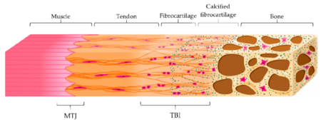
The myotendinous junction (MTJ) is the interface between muscle and tendon, constituting an integrated mechanical unit. The MTJ allows force that is generated by the muscle to be transmitted from the muscle filaments to the collagen fibres of the adjoining tendon. See also about entheses, the connection between tendon/ligament and bone.
Muscle and Tendon/Ligament Structure Overview
Muscles
Synovial joints are acted upon by muscles which are mostly composed of water, proteins, salts, minerals, fat, and carbohydrates. The protein content is made up of various collagens to form their membranes; namely types I, III, IV, and V. The membranes are the endomysium, perimysium, and epimysium.
Sarcomeres are the contractile units and are made up of myosin, actin, and titin. Sarcomeres form myofibres, which are the structural contractile unit of the muscle. Myofibres form muscle fascicles, which are in turn surrounded by perimysium. Muscle fascicles form the whole muscle belly.
Cellularly, a single muscle cell is called a myocyte.
Tendons and Ligaments
Tendons transmit forces between muscles and bones. Ligaments aid in maintaining physiological joint alignment. Tendons and ligaments are made up of water, collagen type I, elastin, and proteoglycans.
The basic structural unit of ligaments and tendons are the tropocollagen molecules that aggregate and form the collagen fibril. Fibrils come together to produce various structures of increasing complexity: sub-fascicles, fascicles, tertiary fibre bundle, and the whole tendon/ligament.
The subunits of tendons are surrounded by endotenon membranes, and that of ligaments by endoligament membranes. The epitenon/epiligament is for the whole tendon/ligament.
Cellularly there are fibroblasts for ligaments, and tenocytes for tendons. These cells are arranged in rows between the collagen fibres.
Myotendinous Junction Structure and Function


The musculoskeletal system as a whole has both hard and soft tissues. The interfaces between these tissues have gradients in order to reduce stress concentrations at the junction sites. The interfaces between tendons/ligaments and bone are called entheses, while the interfaces between tendons and muscles are called myotendinous junctions (MTJ).
The MTJ allows a gradual transition between the stiff tendon and the softer muscle. The main difference to the enthesis, is that the MTJ connects a mostly cellular tissue (the muscle), to a mostly extra-cellular matrix based tissue (the tendon).
Finger-like Processes
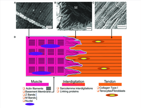
The MTJ has been described as having conical like finger projections that interdigitate with the tendon extracellular matrix with invaginations and evaginations of the sarcolemma (the muscle cell membrane). The finger-like processes might have various functions.
- It increases the surface area by 10-20 times compared to a planar surface, and in doing so decreases stress.
- It also positions the membranes at very low angles relative to the applied stress, and in doing so it causes the membranes to be primarily subjected to shear stress. The adhesion strength is theoretically much higher during shear loading compared to tension loading.
The fingers are not seen in 3D reconstruction, as the fingers are a reflection of the way the tissue is cut. In 3D the structures may be more suitably named ridge-like protrusions and furrow-like indentations. The tendon tissue forms the ridge-like protrusions, and the myofibrils connect to the tendon tissue through these protrusions. The sarcomeres run parallel to the ridge-like protrusions of the tendon.[3] This is in contrast to previous descriptions of the processes being modelled as circular parabaloids.[4]
Tendinous portion
The tendinous portion is made up of multidirectional collagen fibres. In the tendon the force is longitudinal hence orientation is also longitudinal here. However close to the muscle the force can be in many directions. This allows force transmission laterally via the endomysium to adjacent myofibres.
Muscle portion
As the sarcomeres extend towards the MTJ, they appear to change direction, with the longitudinal axis of the sarcomeres becoming parallel with the major axis of the finger-like processes, allowing force transmission via shear as above. There is a final Z-line from the muscle fibre at the MTJ.
Actin myofilaments of the terminal sarcomeres extend from the final Z-line into the plasma membrane to merge with the tendon tissue. It does this by anchoring the muscle cytoskeleton to the tendon extracellular matrix. There are at least two cytoskeletal proteins, vinculin and talin, that are involved in linking the thin filaments to the extracellular structural proteins. There is a separate structural link that traverses the cell membrane of the muscle called integrin, existing within the cell membrane at the MTJ.
On the extracellular side of the MTJ there is a well-developed basement membrane. The basement membrane contains fibronectin, laminin, and type IV collagen. Some of these proteins have an affinity for the tendinous collagen.
Protein Complexes
The MTJ structurally consists of subsarcolemmal, transmembrane, and extracellular protein complexes.
The actin filaments are bundled by actin-binding proteins, and are linked to the sarcolemma through intracellular proteins. There are also transmembrane protein complexes that act to connect the cystoskeletal components to the basement membrane components, as well as proteins that link the basement membrane to the surrounding collagen-rich matrix.
Clinical Applications
Adaptation
The MTJ is a dynamic structure that can adapt to mechanical stimuli. In rats that exercise regularly there are more branches from the finger-like processes compared to non-exercising rats. The angulation of the processes relative to the longitudinal direction also increased.[5]
Aging
With aging we we see shortening of the interdigitations. There is a decrease in contact area between the sarcolemma and extracellular components, but is associated with muscle atrophy.[6]
Injury
Injuries to the MTJ are common. Muscle failure in situ is not associated with separation at the interface between muscle at tendon, but rather in the body of the muscle cells just proximal to the MTJ. [7]
See Also
References
- ↑ Bianchi E, Ruggeri M, Rossi S, Vigani B, Miele D, Bonferoni MC, Sandri G, Ferrari F. Innovative Strategies in Tendon Tissue Engineering. Pharmaceutics. 2021 Jan 11;13(1):89. doi: 10.3390/pharmaceutics13010089. PMID: 33440840; PMCID: PMC7827834.
- ↑ Sensini, Alberto et al. “Tissue Engineering for the Insertions of Tendons and Ligaments: An Overview of Electrospun Biomaterials and Structures.” Frontiers in bioengineering and biotechnology vol. 9 645544. 2 Mar. 2021, doi:10.3389/fbioe.2021.645544
- ↑ Knudsen AB, Larsen M, Mackey AL, Hjort M, Hansen KK, Qvortrup K, Kjaer M, Krogsgaard MR. The human myotendinous junction: an ultrastructural and 3D analysis study. Scand J Med Sci Sports. 2015 Feb;25(1):e116-23. doi: 10.1111/sms.12221. Epub 2014 Apr 10. PMID: 24716465.
- ↑ Tidball JG, Quan DM. Modifications in myotendinous junction structure following denervation. Acta Neuropathol. 1992;84(2):135-40. doi: 10.1007/BF00311385. PMID: 1381858.
- ↑ Kojima H, Sakuma E, Mabuchi Y, Mizutani J, Horiuchi O, Wada I, Horiba M, Yamashita Y, Herbert DC, Soji T, Otsuka T. Ultrastructural changes at the myotendinous junction induced by exercise. J Orthop Sci 2008: 13 (3): 233–239
- ↑ de Palma L, Marinelli M, Bertoni-Freddari C. Involvement of the muscle-tendon junction in skeletal muscle atrophy: an ultrastructural study. Rom J Morphol Embryol 2011: 52 (1): 105–109.
- ↑ Garrett W, Tidball JG. Myotendinous junction: structure, function, and failure. In: Woo SL-Y, Buckwalter JA. (Eds.), Injury and Repair of Musculoskeletal Soft Tissues. American Academy of Orthopedic Surgeons, Park Ridge 1987.
Literature Review
- Reviews from the last 7 years: review articles, free review articles, systematic reviews, meta-analyses, NCBI Bookshelf
- Articles from all years: PubMed search, Google Scholar search.
- TRIP Database: clinical publications about evidence-based medicine.
- Other Wikis: Radiopaedia, Wikipedia Search, Wikipedia I Feel Lucky, Orthobullets,


