Search results
From WikiMSK
- series: increased yield with clinical-radiologic screening criteria. Radiology. 1994 Aug;192(2):481-4. doi: 10.1148/radiology.192.2.8029419. PMID: 8029419. Capitolunate20 KB (2,469 words) - 10:58, 12 May 2024
- extensor tendinopathy with characteristic MR imaging features". Skeletal Radiology (in English). 36 (3): 203–208. doi:10.1007/s00256-006-0238-6. ISSN 0364-23488 KB (948 words) - 10:56, 12 May 2024
- the LSTV was determined. Survey sequences: In New Zealand, some private radiology providers will do a sagittal survey sequence of the entire spine (also17 KB (2,139 words) - 21:01, 12 July 2023
- The journal RadioGraphics (rsna.org) has useful review articles7 members (0 subcategories, 0 files) - 15:38, 8 March 2022
- External Links (section PACS Radiology Systems)Horizon Radiology ARG Radiology SRG Radiology Ascot Radiology Mercy Radiology TRG Imaging Eastmed Radiology Auckland Xray Services Hamilton Radiology Bay Radiology156 bytes (185 words) - 14:04, 28 June 2020
- Spine Imaging (category Radiology)This article is a stub. The primary role of imaging is the identification of undiagnosed systemic disease. Spine imaging has a significant specificity621 bytes (85 words) - 22:11, 2 March 2022
- Dural Ectasia (section Radiology)This article is still missing information. Dural ectasia is a radiological finding characterised by enlargement of the dura in the spinal cord, especially2 KB (255 words) - 16:09, 1 March 2023
- Non-Structural Scoliosis Other: rheumatoid diseases, trauma, bone tumours. Initial radiological investigations should include whole spine radiographs from the cranium31 KB (3,895 words) - 19:29, 11 November 2023
- Therapeutic Injections of the Hip and Groin. Journal of the Belgian Society of Radiology 2017. 101:6. PMID: 30498802. DOI. Full Text. Malhotra, Gunjan; Hansford11 KB (1,600 words) - 20:52, 10 April 2024
- Hip Radiograph (category Radiology)27536615; PMCID: PMC4972716. Sutter et al.. New developments in hip imaging. Radiology 2012. 264:651-67. PMID: 22919039. DOI.4 KB (543 words) - 20:27, 15 April 2022
- al.. Anterior chest wall: frequency of anatomic variations in children. Radiology 1999. 212:837-40. PMID: 10478254. DOI. Donnelly et al.. Asymptomatic, palpable188 bytes (140 words) - 10:46, 2 August 2021
- Hindfoot Radiographs (category Radiology)This article is a stub. The subtalar joint is difficult to evaluate radiographically. Usually a weight-bearing AP or mortise view along with a lateral2 KB (157 words) - 09:59, 3 March 2022
- Horizon Radiology ARG Radiology SRG Radiology Ascot Radiology Mercy Radiology TRG Imaging Eastmed Radiology Auckland Xray Services Hamilton Radiology Bay Radiology2 KB (789 words) - 20:04, 7 April 2023
- Pelvic Radiograph (category Radiology)This article is still missing information. A pelvis x-ray, also known as a pelvis series or pelvis radiograph, is a single x-ray of the pelvis to include5 KB (584 words) - 16:36, 8 May 2021
- guidelines are those produced by the ESSR (European Society of Musculoskeletal Radiology). They have produced a very helpful set of reference documents. ESSR Shoulder676 bytes (80 words) - 14:21, 16 March 2022
- Lumbar Spine MRI (category Radiology)structures. Assess need for other studies Bogduk, Nikolai. Clinical and radiological anatomy of the lumbar spine. Chapter 19. Edinburgh: Elsevier/Churchill10 KB (664 words) - 18:36, 20 October 2022
- Lumbar Spine Radiographs (category Radiology)widespread adoption of posteroanterior projection?. The British journal of radiology 2019. 92:20190386. PMID: 31356113. DOI. Full Text. Assoc Prof Craig Hacking10 KB (1,446 words) - 09:59, 3 March 2022
- Radiation Safety (category Radiology)practice (C1 2018) are the code of practice for diagnostic and interventional radiology. The key concepts are Justication, Optimisation, and Limitation. Justification13 KB (2,104 words) - 07:36, 18 April 2022
- Bursectomy is sometimes done in resistant cases. Deep infrapatellar bursitis | Radiology Reference Article | Radiopaedia.org1 KB (189 words) - 08:22, 13 February 2022
- calcific deposits for rotator cuff calcific tendinitis. (2020) European Radiology. doi:10.1007/s00330-020-06669-0 - Pubmed Luca Maria Sconfienza, Sara Viganò7 KB (1,018 words) - 17:55, 1 March 2022
- development until advanced OA. It is not known whether asymptomatic but radiologically severe FAI should have surgical intervention. At the point of the development34 KB (4,915 words) - 19:20, 11 November 2023
- This article is still missing information. This article reviews the classification, assessment, and management of sternoclavicular instability and pain16 KB (2,448 words) - 18:26, 6 November 2022
- ISSN 1756-185X. PMID 20374315. Check date values in: |date= (help) H Kang. Radiology Illustrated Spine. Springer. 2014 Literature Review Reviews from the last2 KB (240 words) - 21:01, 14 March 2023
- should interpret pain arising from L4/5 and/or L5/S1 using history/exam/radiology, major disability >1 year, degenerative change at above levels Fairbank15 KB (2,146 words) - 05:46, 30 March 2023
- al. MR Imaging Features of Radial Tunnel Syndrome: Initial Experience. Radiology. 2006;240(1):161–8. Rinkel WD et al. Current evidence for effectiveness4 KB (648 words) - 15:17, 11 March 2023
- Osteomalacia (section Radiologic Findings)D. Looser pseudofractures, fissures, narrow radiolucent lines. Loss of radiologic distinctness of vertebral body trabeculae and concavity of vertebral bodies6 KB (675 words) - 12:25, 4 March 2022
- Osgood-Schlatter and Sinding-Larsen-Johansson diseases of the knee". Skeletal Radiology. 18 (3): 193–197. doi:10.1007/BF00360969. ISSN 0364-2348. PMID 26651055 KB (466 words) - 21:34, 7 April 2022
- evaluation of patients with suspected slipping rib syndrome. Skeletal radiology 2019. 48:741-751. PMID: 30612161. DOI. McMahon et al.. Vertical rib plating9 KB (1,315 words) - 20:48, 7 May 2022
- coronal fat sat sequence needs to be specifically requested at certain radiology providers in New Zealand. MRI T2 coronal Dixon and sagittal images showing16 KB (2,230 words) - 17:10, 17 April 2022
- spinal injuries: emphasis on multidetector CT in cervical spine trauma. Radiology. 2012;263(3):645-660. doi:10.1148/radiol.12110526.5 KB (510 words) - 07:04, 7 May 2022
- Sacral meningeal cysts: evaluation with MR imaging. Radiology. 1993 May;187(2):445-8. doi: 10.1148/radiology.187.2.8475288. PMID: 8475288. Marino D, Carluccio12 KB (1,560 words) - 20:13, 15 April 2022
- High-resolution imaging of the musculoskeletal system. Radiology. 1997 Dec;205(3):593-618. doi: 10.1148/radiology.205.3.9393511. PMID: 9393511. Sodhi H, Panitch15 KB (2,061 words) - 11:11, 23 March 2023
- gluteal tendinopathy in greater trochanteric pain syndrome". European Radiology. 17 (7): 1772–1783. doi:10.1007/s00330-006-0485-x. ISSN 0938-7994. Dadour29 KB (3,572 words) - 16:24, 23 April 2022
- Category:Exam
- Manesh 2017 The evolution of the master diagnostician - dhaliwal 2013 Radiologic errors past present and future - Berlin 2014 Using the Chief Complaint4 KB (396 words) - 13:28, 27 April 2022
- postfixed brachial plexus: a review with surgical implications. Surgical and radiologic anatomy : SRA 2010. 32:251-60. PMID: 20087592. DOI. Stecco C, Pirri C44 KB (5,983 words) - 13:58, 16 February 2024
- features of back mice. Skeletal radiology 2018. 47:137-140. PMID: 28914351. DOI. Bogduk, Nikolai. Clinical and radiological anatomy of the lumbar spine. Chapter4 KB (654 words) - 20:12, 15 April 2022
- Petersilge. MR arthrography for evaluation of the acetabular labrum. Skeletal radiology 2001. 30:423-30. PMID: 11479747. DOI. Chopra A; Grainger AJ (2018). "Comparative16 KB (2,524 words) - 07:17, 29 August 2022
- spinal involvement in diffuse idiopathic skeletal hyperostosis (DISH). Radiology 1976. 119:559-68. PMID: 935390. DOI. Utsinger et al.. Diffuse skeletal31 KB (3,839 words) - 09:57, 17 April 2022
- Clinical and Imaging Review. Radiographics : a review publication of the Radiological Society of North America, Inc 2018. 38:1201-1222. PMID: 29995620. DOI15 KB (1,759 words) - 16:50, 10 April 2023
- Wolters Kluwer Health/Lippincott Williams & Wilkins, 2012. Carrying angle | Radiology Reference Article | Radiopaedia.org. Link10 KB (1,415 words) - 16:59, 30 April 2022
- mislead by irrelevant findings. The second caveat is that the reliability of radiology reports is not perfect. The quality is operator dependent and is subject85 KB (12,735 words) - 15:16, 15 April 2023
- rotation, hyperreflexia, dysdiadochokinesia, hypoaesthesia to pinprick Radiological evidence of instability or compression of the neuroaxis is required for4 KB (100 words) - 19:08, 5 May 2022
- under CC BY-NC-SA 3.0 Weidner S, Kellner W, Kellner H. Interventional radiology and the musculoskeletal system. (2004) Best practice & research. Clinical7 KB (951 words) - 07:16, 14 June 2021
- therapists accessed were osteopaths (18%), and chiropractors (28%). Diagnostic radiology was used in 34% of cases and a GP seen by 46% of cases. An orthopaedic12 KB (1,776 words) - 11:22, 4 March 2022

File:Radiology.png (128 × 128 (6 KB)) - 16:54, 3 March 2022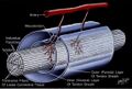
File:Tendon sheath.jpg High-resolution imaging of the musculoskeletal system. Radiology. 1997 Dec;205(3):593-618. doi: 10.1148/radiology.205.3.9393511. PMID: 9393511. This file is copyrighted(1,096 × 740 (119 KB)) - 16:47, 11 August 2021- management. Switzerland: Springer, 2016. Bogduk, Nikolai. Clinical and radiological anatomy of the lumbar spine. Edinburgh: Elsevier/Churchill Livingstone17 KB (2,599 words) - 15:17, 11 March 2023
- for both axial and peripheral SpA. Sacroiliitis on MRI is an important radiological finding in classification. The term spondyloarthritis is preferred over10 KB (868 words) - 21:01, 14 March 2023
- Thoracic zygapophysial joint pain: Complete relief of pain on selective radiologically controlled intra-articular anaesthesia of the targeted joint followed10 KB (1,498 words) - 21:03, 18 March 2022
- Clinical and Imaging Review. Radiographics : a review publication of the Radiological Society of North America, Inc 2018. 38:1201-1222. PMID: 29995620. DOI4 KB (144 words) - 20:36, 11 March 2023
- first sign of avascular necrosis is slight sclerosis. See radiopaedia for radiology cases. Initial radiographs can miss 5-20% of fractures in the acute setting14 KB (1,852 words) - 07:38, 18 April 2022
- Lifestyle Neurology Paediatrics Pain Maps Pharmacology Physiology Psychiatry Radiology Rheumatology Tendinopathies Autism and Chronic Pain CRPS Dysautonomia Myofascial22 members (17 subcategories, 0 files) - 19:08, 4 August 2020
- Physiology Precision Techniques Presenting Complaints Procedures Psychiatry Radiology Regional Conditions Rheumatology Statistics Tendinopathies29 members (29 subcategories, 0 files) - 06:20, 2 April 2022
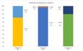
File:Vertebral Arrangement Patterns.jpg spinal segment variants on numbering vertebral levels at lumbar MR imaging. Radiology. 2011 Apr;259(1):196-202. doi: 10.1148/radiol.11081511. PMID: 21436097(1,313 × 812 (69 KB)) - 19:56, 6 January 2022- Redlund-Johnell, Inga (1986-01). "The costoclavicular joint". Skeletal Radiology. 15 (1): 25–26. doi:10.1007/bf00355069. ISSN 0364-2348. Check date values18 KB (2,468 words) - 20:49, 7 May 2022

File:Hip injection anterior longitudinal approach.jpg Therapeutic Injections of the Hip and Groin. Journal of the Belgian Society of Radiology 2017. 101:6. PMID: 30498802. DOI. Full Text. This work is licensed under(400 × 626 (35 KB)) - 14:51, 21 June 2021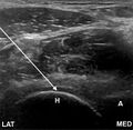
File:Hip joint injection anterolateral approach ultrasound.jpg Therapeutic Injections of the Hip and Groin. Journal of the Belgian Society of Radiology 2017. 101:6. PMID: 30498802. DOI. Full Text. This work is licensed under(640 × 626 (56 KB)) - 14:59, 21 June 2021- Pain 2009. 147:17-9. PMID: 19762151. DOI. Bogduk, Nikolai. Clinical and radiological anatomy of the lumbar spine. Chapter 15. Edinburgh: Elsevier/Churchill31 KB (4,193 words) - 15:21, 11 March 2023
- radicular pain of the buttock and leg. Higher nerve roots can be involved. Radiological diagnosis is difficult. With conventional imaging the area of interest5 KB (778 words) - 09:59, 17 April 2022
- Tucker S, Taylor BA. Orientation of lumbar pars defects: implications for radiological detection and surgical management. J Bone Joint Surg Br. 1998 Mar;80(2):208-118 KB (1,203 words) - 20:36, 6 May 2022
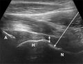
File:Hip joint injection anterior longitudinal approach ultrasound.jpg Therapeutic Injections of the Hip and Groin. Journal of the Belgian Society of Radiology 2017. 101:6. PMID: 30498802. DOI. Full Text. This work is licensed under(640 × 491 (48 KB)) - 14:59, 21 June 2021- test of either eye, abnormal sialometry or Saxon test for the glands, or radiological evidence such as on MRI, CT, or US. Serological or histological findings:15 KB (1,511 words) - 18:34, 12 March 2023
- dorsi Review by Poilliot et al, free access Bogduk, Nikolai. Clinical and radiological anatomy of the lumbar spine. Edinburgh: Elsevier/Churchill Livingstone16 KB (1,579 words) - 14:36, 29 May 2022

File:Hip AP view radiographic signs.png Zanetti, M., & Pfirrmann, C. W. A. (2012). New Developments in Hip Imaging. Radiology, 264(3), 651–667. doi:10.1148/radiol.12110357 This file is copyrighted(1,039 × 260 (129 KB)) - 22:21, 22 June 2021- "Magnetic resonance imaging of pyomyositis in 43 cases". European Journal of Radiology. 35 (1): 59–64. doi:10.1016/s0720-048x(99)00108-4. ISSN 0720-048X. Check29 KB (3,588 words) - 18:14, 12 March 2023
- Pain Disorder Rare Diseases Sodium Channelopathies Radiology Select [►] to view subcategories Radiology Hindfoot Radiographs Hip Radiograph Lumbar Spine5 KB (863 words) - 18:15, 12 March 2023
- Baxter's neuropathy in recalcitrant heel pain syndrome: part I". Surgical and radiologic anatomy: SRA. 41 (1): 29–41. doi:10.1007/s00276-018-2124-z. ISSN 1279-85178 KB (887 words) - 15:17, 11 March 2023
- 8.84, and negative LR was 0.17. Bogduk. Instability In: Clinical and radiological anatomy of the lumbar spine. Elsevier 2012 Weiler PJ, King GJ, Gertzbein11 KB (1,514 words) - 11:27, 13 September 2021
- date values in: |date= (help) Bogduk. Low back pain In: Clinical and Radiological Anatomy of the Lumbar Spine. 5th Edition. Elsevier 2012 Literature Review2 KB (318 words) - 21:30, 17 April 2022
- Spine Society 2005. 5:615-22. PMID: 16291100. DOI. Kitab et al.. Anatomic radiological variations in developmental lumbar spinal stenosis: a prospective, control-matched6 KB (966 words) - 09:50, 17 April 2022
- sustained reapplied loads in the usual way. Bogduk, Nikolai. Clinical and radiological anatomy of the lumbar spine. Chapter 7. Edinburgh: Elsevier/Churchill12 KB (1,589 words) - 16:59, 30 April 2022
- muscle for investigation of neurogenic thoracic outlet syndrome". Skeletal Radiology. 38 (11): 1083–1087. doi:10.1007/s00256-009-0714-x. ISSN 1432-2161. PMID 1944070533 KB (4,365 words) - 20:32, 11 March 2023
- Nicholas; Smith, Toby (2014-03). "Diagnostic test accuracy of clinical and radiological assessments for medial patella plica syndrome: a systematic review and10 KB (1,394 words) - 22:57, 25 April 2022
- 2-year follow-up considering features of injury mechanism and somatic, radiologic, and psychosocial findings. Medicine 1995. 74:281-97. PMID: 7565068. DOI19 KB (3,177 words) - 09:47, 3 March 2022
- dislocations / subluxations 2. Kyphoscoliosis 3. Skin hyperextensibility 3. Radiologically mild osteopenia 4. Tissue fragility, including atrophic scars 5. Easy26 KB (1,610 words) - 19:25, 16 December 2022

File:Berlin2014 Radiologic errors past present and future.pdf File uploaded with MsUpload(250 KB) - 08:24, 13 September 2020- Lifestyle Neurology Paediatrics Pain Maps Pharmacology Physiology Psychiatry Radiology Rheumatology Tendinopathies Autism and Chronic Pain CRPS Dysautonomia Myofascial4 KB (609 words) - 11:32, 2 April 2022
- are study notes taken from Chapter 8 of: Bogduk, Nikolai. Clinical and radiological anatomy of the lumbar spine. Edinburgh: Elsevier/Churchill Livingstone15 KB (2,241 words) - 17:34, 30 April 2022
- are study notes taken from Chapter 13 of: Bogduk, Nikolai. Clinical and radiological anatomy of the lumbar spine. Edinburgh: Elsevier/Churchill Livingstone14 KB (530 words) - 17:33, 30 April 2022
- L4/L5 level. Be aware that the sides are reversed compared to standard radiological imaging. There is only one negative RCT for the treatment of lumbar facet15 KB (1,582 words) - 14:14, 13 September 2021
- internal disc disruption. Importantly, there is no correlation between radiological signs on plain films or CT of osteoarthritis and the joint being painful41 KB (6,142 words) - 18:31, 3 November 2022
- instability of the axis or spinal cord injury. In less severe trauma, there is radiological evidence that the alar and transverse ligaments can be injured in whiplash30 KB (4,725 words) - 21:00, 18 March 2022
- Epub 2011 Jan 25. PMID: 21266006. Bogduk. Low back pain In: Clinical and Radiological Anatomy of the Lumbar Spine. 5th Edition. Elsevier 2012 Literature Review15 KB (2,219 words) - 21:55, 18 March 2022
- Inability to identify anatomic landmarks Severe to morbid obesity blocking radiologic (fluoroscopic) visualisation Deformity of sacral coccygeal area secondary14 KB (1,977 words) - 19:21, 22 January 2023
- Diagnosis and Classification. Radiographics : a review publication of the Radiological Society of North America, Inc 2015. 35:765-79. PMID: 25969933. DOI.19 KB (2,757 words) - 06:02, 2 April 2022
- Disruption Sacroiliac Joint Pain Bogduk. Low back pain In: Clinical and Radiological Anatomy of the Lumbar Spine. 5th Edition. Elsevier 2012 Kalichman L,18 KB (2,446 words) - 09:52, 30 July 2022
- imaging or CT on treatment and outcome--multicenter randomized trial. Radiology. 2004 May;231(2):343-51. doi: 10.1148/radiol.2312030886. Epub 2004 Mar59 KB (8,684 words) - 10:49, 7 May 2022
- only examine the immediate effects of US and that there was no apparent radiological standardisation of the lesion (e.g., size, location within muscle) being20 KB (2,897 words) - 20:23, 2 August 2022
- rate of false negatives Cauda equina syndrome is a clinical rather than radiological diagnosis, but MRI can inform which patients with the clinical syndrome21 KB (2,900 words) - 15:21, 11 March 2023
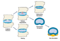
File:Endplate fracture consequences.png Own work. Based off Bogduk. Clinical and Radiological Anatomy of the Lumbar Spine. Fifth Edition. 2012 This work is licensed under the Creative Commons(1,920 × 1,309 (114 KB)) - 19:10, 16 September 2021- horn of the hyoid bone: a landmark for cervical surgery". Surgical and Radiologic Anatomy. 27 (1): 33–36. doi:10.1007/s00276-004-0263-x. ISSN 0930-103825 KB (3,213 words) - 20:21, 7 May 2022
- 2-year follow-up considering features of injury mechanism and somatic, radiologic, and psychosocial findings. Medicine 1995. 74:281-97. PMID: 7565068. DOI25 KB (4,224 words) - 19:20, 7 January 2023
- are study notes taken from Chapter 10 of: Bogduk, Nikolai. Clinical and radiological anatomy of the lumbar spine. Edinburgh: Elsevier/Churchill Livingstone28 KB (4,039 words) - 17:34, 30 April 2022
- useful as a screening test. Many patients with significant and symptomatic radiological changes do not have a positive compression test. Shoulder abduction relief41 KB (5,539 words) - 15:21, 11 March 2023
- principal compression trabeculae are visible. (Drawing modified from A Radiological Study on the Trabecular Pattern in the Upper End of the Femur in Post-Menopausal28 KB (3,720 words) - 05:24, 13 March 2023
- suspected pathologic site that correlates with the patient's clinical and radiological picture. If the patient is on anticoagulants the doctor should weigh30 KB (4,581 words) - 20:40, 22 March 2023
- Disease-specific: Involved knee: right 43.5%, left 56.5% Kellgren-Lawrence radiologic OA assessment grade (out of 4, higher indicates more evidence of disease7 KB (996 words) - 12:23, 25 March 2022


