Search results
From WikiMSK
- Transitional Vertebral Anatomy (category Lumbar Spine Anatomy) (section Lumbosacral Transitional Anatomy)transitional anatomy but the total thoracolumbosacral count remains the same. It is important to accurately describe transitional anatomy as failing to17 KB (2,139 words) - 21:01, 12 July 2023
- and pelvis innervation. This article is a general overview of spinal cord anatomy. See nociception and basic neurophysiology for a more detailed overview15 KB (1,759 words) - 16:50, 10 April 2023
- Ilioinguinal Neuralgia (section Anatomy)This article is a stub. Main article: Ilioinguinal Nerve The ilioinguinal nerve originates from the L1 nerve root along with the iliohypogastric nerve2 KB (159 words) - 20:59, 16 May 2023
- Sensory Polyneuropathies (section Anatomy)This article is still missing information. The terms "polyneuropathy," "peripheral neuropathy," and "neuropathy" are often often used interchangeably but14 KB (961 words) - 19:27, 27 November 2023
- Iliotibial Band Syndrome (section Anatomy)This article is still missing information. Iliotibial band syndrome is a common cause of lateral knee pain in runners. The ITB is a thickened part of the10 KB (1,592 words) - 21:16, 15 April 2022
- update - 1.03 MB (f) Tamborrini 2017 - The Rotator Interval – A Link Between Anatomy and Ultrasound - 1.44 MB (f) Sambandam 2015 - Rotator cuff tears: An evidence2 KB (226 words) - 11:49, 28 August 2023
- Small Fibre Neuropathy (section Nerve Anatomy)Written by: Dr Jeremy Steinberg – created: 24 March 2021; last modified: 20 May 2023 This article is still missing information. Small fiber neuropathy29 KB (3,612 words) - 21:12, 20 May 2023
- Coccydynia (section Anatomy)rigid coccyx. The optimal management remains unknown. Main article: Coccyx Anatomy The coccyx has between three to five vertebrae. Four segments is the most18 KB (2,612 words) - 20:13, 8 November 2023
- Radiocarpal Joint (category Hand and Wrist Anatomy)This article is a stub. The radiocarpal joint is formed by the articulation between the distal end of the radius, a bone in the forearm, and the proximal3 KB (221 words) - 21:44, 26 March 2023
- Dermatomes (category Spine Anatomy)can cause an atypical presentation of sciatica. Transitional lumbosacral anatomy may result in the L4 nerve root serving the usual function of L5 in L5 sacralisation44 KB (5,983 words) - 13:58, 16 February 2024
- attaches to the tibia, malleoli, and talus. Ankle Bony Anatomy – Anterior View Ankle and Hindfoot Bony Anatomy – Lateral View There are three groups of ligaments5 KB (663 words) - 07:31, 8 May 2022
- 6 members (6 subcategories, 0 files) - 11:33, 3 August 2020
- Hip Joint Injection (section Anatomy)This article is still missing information. Hip joint injections can be performed with a variety of image guidance, including fluoroscopy and ultrasound11 KB (1,600 words) - 20:52, 10 April 2024
- This article is still missing information. Isolated scaphotrapeziotrapezoid (STT) joint osteoarthritis is a fairly common cause of radial sided wrist pain4 KB (693 words) - 20:23, 22 March 2022
- This article is still missing information. The Ilioinguinal and iliohypogastric nerve injection, or nerve block, is a valuable diagnostic, prognostic,7 KB (905 words) - 21:54, 16 May 2023
- This article is still missing information. The coccyx (plural: coccyges) is the series of rudimentary vertebrae forming the caudal termination of the vertebral6 KB (768 words) - 17:27, 30 April 2022
- Lumbar Zygapophyseal (Facet) Joint (category Lumbar Spine Anatomy)This article is a stub.386 bytes (5 words) - 05:32, 24 March 2023
- Cervical Zygapophyseal (Facet) Joint (category Cervical Spine Anatomy)C0-1 C1-2 C2-7 Anatomy 1. Superior facets of C0 (Atlas): 28° in sagittal and transverse planes214 bytes (5 words) - 08:47, 12 June 2022
- Cervical Spine Biomechanics (category Cervical Spine Anatomy)C0-1 C1-2 C2-7 Anatomy 1. Superior facets of C0 (Atlas): 28° in sagittal and transverse planes 2. No disc 1. C1 has convex facet joint surface (allow646 bytes (50 words) - 08:49, 12 June 2022
- Supraorbital Nerve Injection (section Anatomy)This article is still missing information. The supraorbital nerve is a branch of the terminal cutaneous branches of the frontal nerve. It runs through2 KB (307 words) - 11:31, 4 March 2022
- and Instability Sternoclavicular Joint Injection Epperson & Varacallo. Anatomy, Shoulder and Upper Limb, Sternoclavicular Joint. 2021. . PMID: 307259437 KB (586 words) - 11:33, 8 May 2022
- This article is a stub. There is surprisingly little consensus on the anatomy of the lateral hip rotators Lumbar Plexus Subcostal nerve Iliohypogastric658 bytes (106 words) - 17:35, 4 April 2022
- This article is still missing information. The hip joint is a synovial joint between the femoral head and the acetabulum of the pelvis. The hip is a ball13 KB (1,660 words) - 16:51, 30 April 2022
- Distal Clavicle Osteolysis (section Anatomy)Gómez CM, Umpire DF, Pathria MN. Imaging of the Acromioclavicular Joint: Anatomy, Function, Pathologic Features, and Treatment. Radiographics. 2020 Sep-Oct;40(5):1355-13825 KB (656 words) - 11:17, 7 February 2024
- Interspinous Oedema (section Anatomy)some cases, most commonly in women aged between 15-35. As outlined in the anatomy section, these ligaments do not provide significant resistance to forward16 KB (2,230 words) - 17:10, 17 April 2022
- Superior Cluneal Nerve Injection (section Anatomy)This page or section deals with a topic that is not widely recognised or accepted. Please use your clinical judgement and note that this is not necessarily3 KB (371 words) - 15:09, 6 June 2021
- This article is still missing information. The talocalcaneal joint, also called the clinical subtalar joint, is an important and complex joint in the hindfoot9 KB (1,291 words) - 07:57, 8 May 2022
- Cervical Intervertebral Discs (category Cervical Spine Anatomy)C0-1 C1-2 C2-7 Anatomy 1. Superior facets of C0 (Atlas): 28° in sagittal and transverse planes5 KB (394 words) - 08:52, 12 June 2022
- Greater Occipital Nerve Injection (section Anatomy)This article is still missing information. The greater occipital nerve is a popular target for treating and preventing headache disorders. The greater5 KB (755 words) - 20:31, 15 March 2022
- Adult Acquired Flatfoot Deformity (section Anatomy)the foot with continued progressive deformity of the foot and ankle. The anatomy of the foot and ankle are complex, with multiple structures involved in19 KB (2,657 words) - 11:22, 11 September 2023
- and pain. See Sternoclavicular Joint Anatomy for a review of the anatomy See also: Sternoclavicular Joint Anatomy The Sternoclavicular joint (SCJ) is inherently16 KB (2,448 words) - 18:26, 6 November 2022
- Ligaments of the Lumbar Spine (category Lumbar Spine Anatomy)This article is a stub. There are three parts to the interspinous ligaments. Ventrally there are fibres that run posterocranially from the dorsal aspect3 KB (515 words) - 17:32, 30 April 2022
- Sacroiliac Joint (redirect from Sacroiliac Joint Anatomy)Poilliot et al, free access Bogduk, Nikolai. Clinical and radiological anatomy of the lumbar spine. Edinburgh: Elsevier/Churchill Livingstone, 2012 Poilliot16 KB (1,579 words) - 14:36, 29 May 2022
- Dupuytren Disease (section Anatomy)contracture. This is called Dupuytren contracture The anatomy of the hand is very complex as is the anatomy of which structures are involved in Dupuytren disease10 KB (1,475 words) - 10:22, 17 April 2022
- Os Peroneum and Os Peroneum Syndrome (section Anatomy)Os peroneum (OP) normal anatomy. Peroneal tendons (peroneus brevis 1, peroneus longus 2) travel along the lateral surface of the calcaneus that presents9 KB (1,406 words) - 19:55, 7 March 2022
- This article is still missing information. Pain following hernia surgery is common, generally subsiding within approximately two months. However, a proportion9 KB (1,156 words) - 21:55, 16 May 2023
- Slipping Rib Syndrome (section Anatomy)Slipped rib syndrome anatomy Slipping Rib Syndrome Epidemiology Any age, females > males Pathophysiology loss of fibrous or cartilaginous attachments between9 KB (1,315 words) - 20:48, 7 May 2022
- Sacroiliac Joint Injection (section Anatomy)precision treatments for this structure. Main article: Sacroiliac Joint Anatomy The sacroiliac joint is around 1-2mm wide and is formed within S1, S2, and9 KB (1,317 words) - 20:12, 28 April 2022
- Superior Cluneal Nerve Entrapment (section Anatomy)Switzerland: Springer, 2016. Bogduk, Nikolai. Clinical and radiological anatomy of the lumbar spine. Edinburgh: Elsevier/Churchill Livingstone, 2012. Maigne17 KB (2,599 words) - 15:17, 11 March 2023
- Lumbar Spine Bone Metabolic Activity (category Lumbar Spine Anatomy)This article is a stub. Lumbar Spine Shape and Metabolic Activity - Toor 2020 - 1.77 MB (f)186 bytes (17 words) - 17:34, 30 April 2022
- Thoracolumbar Fascia (category Lumbar Spine Anatomy)Willard et al.. The thoracolumbar fascia: anatomy, function and clinical considerations. Journal of anatomy 2012. 221:507-36. PMID: 22630613. DOI. Full235 bytes (45 words) - 17:35, 30 April 2022
- Chronic Post-Traumatic Neck Pain (section Anatomy)pathology is James Taylor's The Cervical Spine: An atlas of normal anatomy and the morbid anatomy of ageing and injuries. The zygaphophyseal joints are an important19 KB (3,177 words) - 09:47, 3 March 2022
- Finger Anatomy (category Hand and Wrist Anatomy)Konstantinou P, Pinto I, Karavelis A, Kostretzis L (2017) Extensor Mechanism’s Anatomy at the Metacarpophalangeal Joint. MOJ Orthop Rheumatol 8(4): 00319. DOI:7 KB (936 words) - 17:20, 30 April 2022
- see effect in <10 min Bleeding/hematoma Infection Intravascular injection Anatomy for EM - Fascia Iliaca Block Video: Fascia Iliaca Block2 KB (265 words) - 21:51, 4 May 2022
- Lateral Hip Sonoanatomy (category Pelvis, Hip, and Thigh Anatomy)This article is a stub.114 bytes (5 words) - 19:46, 11 April 2022
- Suprascapular Nerve Injection (section Anatomy)Adhesive Capsulitis, calcific tendinitis, and bursitis. Shoulder Shoulder Anatomy Shoulder Conditions Shoulder Procedures Shoulder Examination Shoulder Pain788 bytes (98 words) - 14:20, 23 August 2020
- Lumbar Spine Sonoanatomy (category Lumbar Spine Anatomy)This article is a stub.140 bytes (5 words) - 21:15, 2 March 2022
- Diaphragm (category Chest Wall Anatomy)This article is a stub. Diaphragm two muscles - Pickering 2002 - 197 KB (f)775 bytes (13 words) - 08:22, 8 May 2022
- Cervical Facet Joint Injection (section Anatomy)This article is a stub. Literature Review Reviews from the last 7 years: review articles, free review articles, systematic reviews, meta-analyses, NCBI559 bytes (51 words) - 15:25, 8 May 2021
- Genicular Nerve Injection (section Anatomy)steroid if using. Knee and Leg Knee and Leg Anatomy Knee and Leg Conditions Knee and Leg Procedures Lower Limb Anatomy Acute Knee Pain Calf Pain Differential2 KB (299 words) - 15:11, 8 May 2021
- Sternoclavicular Joint Injection (section Anatomy). The topics of sternoclavicular joint pain and sternoclavicular joint anatomy are addressed elsewhere. The sternoclavicular joint has a small meniscus4 KB (536 words) - 15:01, 25 April 2021
- Superficial Radial Nerve (section Anatomy)part of the sensory innervation of the hand. This article explores the anatomy, function, and clinical significance of the superficial radial nerve in3 KB (356 words) - 17:37, 15 March 2023
- Supratrochlear Nerve Injection (section Anatomy)This article is still missing information. The supratrochlear nerve is a terminal cutaneous branch of the frontal nerve. The frontal nerve arises from2 KB (248 words) - 07:35, 7 March 2022
- Elbow Joint (category Elbow and Forearm Anatomy)This article is a stub.585 bytes (5 words) - 17:09, 30 April 2022
- First Carpometacarpal Joint (Trapeziometacarpal Joint) (category Hand and Wrist Anatomy)This article is a stub.625 bytes (5 words) - 17:20, 30 April 2022
- Sacrum (section Gross Anatomy)L5, fusion of the coccyx, and spina bifida (see Transitional Vertebral Anatomy). The sacrum is wedge shaped with concave anterior and convex posterior4 KB (524 words) - 17:29, 30 April 2022
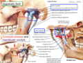
- TMJ Examination (section Anatomy)This article is still missing information. Oral exam Head/chin deviations C curve occurs due to hypomobility, the mandible deviates to the involved side3 KB (240 words) - 11:30, 4 March 2022
- Lesser Occipital Nerve Injection (section Anatomy)This article is still missing information. The lesser occipital nerve (LON) is commonly injected alongside the greater occipital nerve for headache disorders1 KB (196 words) - 20:30, 15 March 2022
- Glenohumeral Joint (category Shoulder Anatomy)This article is a stub.1 KB (5 words) - 17:21, 30 April 2022
- Auriculotemporal Nerve Injection (section Anatomy)This article is still missing information. The auriculotemporal nerve is a branch of the mandibular nerve (V3) that runs with the superficial temporal2 KB (295 words) - 17:40, 7 March 2022
- Paraspinous Cervical Block (section Anatomy)This page or section deals with a topic that is not widely recognised or accepted. Please use your clinical judgement and note that this is not necessarily2 KB (332 words) - 16:43, 8 May 2021
- Plantar Plate Injury (Turf Toe) (section Anatomy)This article is still missing information. See Drakos et al for a brief review. First MTPJ integrity is related primarily to the joint capsule, collateral3 KB (538 words) - 19:50, 26 March 2022
- Carpal Tunnel Injection (section Anatomy)This article is still missing information. The flexor retinaculum attaches to four areas. Namely the pisoform, the scaphoid, the hook of the hamate, and3 KB (441 words) - 05:47, 22 January 2022
- presence of pain see Pain Oriented Sensory Testing. Main article: Spinal Cord Anatomy Part or all of this article or section is derived from Numbness by ddxof5 KB (595 words) - 11:17, 7 May 2022
- Third Occipital Nerve Injection (section Anatomy)nearly reach the superior nuchal line. See Kim et al for a review of the anatomy The third occipital nerve can be blocked using ultrasound guidance, which3 KB (518 words) - 20:31, 15 March 2022
- sensory disturbance at levels not explained by standard imaging. Transitional anatomy if present, can also contribute to variable clinical presentations. Diagnostic3 KB (523 words) - 17:32, 30 April 2022
- for understanding the behavior of biological tissues in human and animal anatomy. These properties determine how tissues respond to different types of stress9 KB (1,290 words) - 07:54, 24 March 2023
- Knee Joint Injection (section Anatomy)This article is a stub. Infection, uncontrolled coagulopathy, joint prosthesis, poor response to previous injections. Allergy to eggs or feathers is a3 KB (494 words) - 16:44, 23 March 2023
- De Quervain Injection (section Anatomy)This article is still missing information. Injection for De Quervain Tendinopathy. The APL and EPB usually run together in the first dorsal compartment4 KB (496 words) - 14:21, 16 March 2022
- Peroneal Tendon Sheath Injection (section Anatomy)This article is still missing information. The peroneal tendons are located in the lateral compartment of the leg behind the lateral malleolus. Peroneus3 KB (456 words) - 17:55, 2 June 2021
- Ankle Joint Injection (section Anatomy)This article is still missing information. Main article: Ankle Joint Anatomy The ankle joint, or tibiotalar joint, is a hinged synovial joint. It is created4 KB (554 words) - 19:42, 4 June 2021
- Sacroiliac Joint Pain (section Anatomy)muscles, motor control, and alignment. Main article: Sacroiliac Joint Anatomy The sacroiliac joint is a diarthrodial synovial joint, and only the anterior26 KB (4,171 words) - 21:01, 14 March 2023
- Hoffa's Fat Pad Pain (section Anatomy and Physiology)This article is still missing information. The infrapatellar fat pad (figure 1) also known as Hoffa's fat pad is the primary adipose structure within the4 KB (628 words) - 08:20, 13 February 2022
- Acromioclavicular Joint Injection (section Anatomy)This article is still missing information. The ACJ is a small joint therefore the injected volume should reflect this i.e maximum 2mL. pain - arthropathy4 KB (579 words) - 11:22, 4 March 2022
- Inferior Cluneal Nerve Entrapment (section Anatomy)This article is a stub. The cluneal nerves are a group of pure sensory nerves (superior, medial or middle, and inferior) that provide sensory supply to3 KB (318 words) - 15:17, 11 March 2023
- Quadratus Femoris Injection (section Anatomy)This article is a stub. The ischiofemoral space is bounded by the ischial tuberosity medially, the lesser trochanter of the femur laterally, the femoral5 KB (536 words) - 11:46, 17 April 2023
- Proximal Radioulnar Joint (category Elbow and Forearm Anatomy)This article is a stub. The proximal radioulnar joint is a pivot type synovial joint between the circumference of the head of the radius and the ring formed1 KB (62 words) - 17:10, 30 April 2022
- Sagittal Band Injuries (section Anatomy)repetitive microtrauma, and typically occur in boxers. Main article: Finger Anatomy The extensor hood stabilises the extensor tendon at the MCPJ, whereas the6 KB (680 words) - 10:06, 17 April 2022
- Plantar Fasciitis (section Anatomy)This article is still missing information. Plantar fasciitis is the most common cause of chronic heel pain, with a lifetime prevalence of up to 10%. See4 KB (598 words) - 07:20, 8 May 2022
- Lumbar Intervertebral Discs (category Lumbar Spine Anatomy)This article is a stub. Posterior Disc: sinuvertebral nerve from ventral ramus Anterolateral disc: Grey ramus communicans of the lumbar sympathetic trunk3 KB (72 words) - 08:54, 12 June 2022
- Nerves of the Cervical Spine (category Cervical Spine Anatomy)This article is a stub. The C1 spinal nerve is unique. It does not have a typical dorsal root ganglion. The ganglion cells are located in the rootlets2 KB (322 words) - 21:15, 4 May 2022
- This article is still missing information. The Scaphotrapeziotrapezoid (STT) or triscaphe joint is the joint that is shared between the scaphoid, trapezium5 KB (711 words) - 21:31, 14 March 2023
- (meralgia paraesthetica). Full discussion of the clinical syndrome and the anatomy are discussed elsewhere (See Lateral Femoral Cutaneous Nerve Entrapment9 KB (1,223 words) - 08:54, 15 June 2021
- to steroid leaking into the surrounding soft tissues Shoulder Shoulder Anatomy Shoulder Conditions Shoulder Procedures Shoulder Examination Shoulder Pain4 KB (610 words) - 15:09, 14 June 2021
- Tarsal Tunnel Syndrome (section Anatomy)This article is still missing information. Tarsal tunnel syndrome is an mononeuropathy of the posterior tibial nerve or of one of its branches in the tarsal5 KB (698 words) - 15:37, 11 March 2023
- Cervical Spine Injuries (Acute) (section Anatomy)This is ported content from ddxof It is subject to the compatible CC-BY-SA license. Atlanto-occipital dislocation , atlantoaxial dislocations , potentially5 KB (510 words) - 07:04, 7 May 2022
- This article is still missing information. TMC joint injection may be indicated with failure of first line treatments to treat chronic pain e.g. fromosteoarthritis4 KB (635 words) - 11:24, 4 March 2022
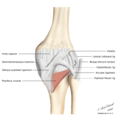
- This article is still missing information. The elbow joint functions as a fulcrum for the forearm and helps position the hand in space. The elbow joint10 KB (1,415 words) - 16:59, 30 April 2022
- Acromioclavicular Joint (category Shoulder Anatomy)This article is a stub. The acromioclavicular joint (ACJ) is formed by the junction between the acromion process of the scapula and the distal clavicle2 KB (124 words) - 17:21, 30 April 2022
- Distal Radioulnar Joint (category Hand and Wrist Anatomy)This article is a stub. The distal radioulnar joint is a pivot type synovial joint existing between the ulnar notch of the distal radius and the head of1 KB (81 words) - 17:19, 30 April 2022
- Metatarsophalangeal Joints (category Foot and Ankle Anatomy)This article is still missing information. The five metatarsophalangeal joints are each composed of a convex metatarsal head and a concave proximal phalanx3 KB (289 words) - 16:55, 30 April 2022
- Baxter's Nerve Entrapment (section Anatomy)abductor hallucis muscle and quadratus plantae. The Gray's anatomy image and many other anatomy textbook images are incorrect. The Baxter nerve is quite8 KB (887 words) - 15:17, 11 March 2023
- Lateral Plantar Nerve Entrapment (section Anatomy)Written by: Dr Jeremy Steinberg – created: 16 April 2022; last modified: 11 March 2023 This article is still missing information. Lateral plantar nerve5 KB (631 words) - 15:17, 11 March 2023
- This article is still missing information. Quadrilateral Space Syndrome (QSS) is a relatively uncommon disorder, initially described by Cahill and Palmer3 KB (456 words) - 09:27, 24 March 2023
- Greater Trochanteric Bursa Injection (section Anatomy)This article is still missing information. Greater femoral trochanteric bursa injections under ultrasound-guidance ensures the injectate is accurately4 KB (592 words) - 11:10, 4 March 2022
- Atlanto-occipital Joint (category Cervical Spine Anatomy)C0-1 C1-2 C2-7 Anatomy 1. Superior facets of C0 (Atlas): 28° in sagittal and transverse planes3 KB (284 words) - 08:48, 12 June 2022
- Trigger Finger (section Anatomy and Pathophysiology)Konstantinou P, Pinto I, Karavelis A, Kostretzis L (2017) Extensor Mechanism’s Anatomy at the Metacarpophalangeal Joint. MOJ Orthop Rheumatol 8(4): 00319. DOI:6 KB (921 words) - 20:13, 15 April 2022
- Metacarpophalangeal Joint (category Hand and Wrist Anatomy)This article is a stub. The metacarpophalangeal joints (MCP) are condyloid joints situated between the metacarpal bones and the proximal phalanges of the1 KB (136 words) - 17:20, 30 April 2022
- Neuronopathies (section Anatomy)This article is still missing information. Neuronopathy means the cell body is affected rather than the myelin or axon as in peripheral neuropathy. This9 KB (780 words) - 20:59, 27 November 2023
- Interphalangeal Joints (Foot) (category Foot and Ankle Anatomy)This article is a stub. The great toe (hallux) has a single interphalangeal joint. The lateral four times have both a proximal and distal interphalangeal987 bytes (107 words) - 16:54, 30 April 2022
- Hip Examination (section Anatomy)This article is still missing information. Think of the hip with it's muscles as the rotator cuff of the lower limbs. Action: Hip abductors – 3 Glut medius7 KB (1,139 words) - 21:08, 4 May 2022
- Sacral Insufficiency Fracture (section Anatomy)This article is still missing information. Sacral insufficiency fractures are a relatively common fracture in elderly women that is notoriously difficult8 KB (1,142 words) - 19:08, 17 November 2022
- Superior Gluteal Nerve Entrapment (section Anatomy)Written by: Dr Jeremy Steinberg – created: 6 June 2021; last modified: 11 March 2023 This article is still missing information. This page or section deals6 KB (874 words) - 15:17, 11 March 2023
- Spondylolysis (section Anatomy)This article is still missing information. Spondylolysis, also known as a pars defect, is a unilateral or bilateral fracture in the pars interarticularis8 KB (1,203 words) - 20:36, 6 May 2022
- Intertarsal and Tarsometatarsal Joints (category Foot and Ankle Anatomy)This article is still missing information. The intertarsal joints include the three cuneonavicular joints, the intercuneiform joints, the cuboideonavicular3 KB (348 words) - 16:55, 30 April 2022
- Ganglion Impar Injection (section Anatomy)technique and may provide better results than blockade. Main article: Coccyx Anatomy The ganglion impair is a midline sympathetic ganglion anterior to the upper7 KB (988 words) - 19:21, 22 January 2023
- Lumbar Instability (section Anatomy)negative LR was 0.17. Bogduk. Instability In: Clinical and radiological anatomy of the lumbar spine. Elsevier 2012 Weiler PJ, King GJ, Gertzbein SD. Analysis11 KB (1,514 words) - 11:27, 13 September 2021
- Atlanto-axial Joint (category Cervical Spine Anatomy)C0-1 C1-2 C2-7 Anatomy 1. Superior facets of C0 (Atlas): 28° in sagittal and transverse planes6 KB (744 words) - 08:48, 12 June 2022
- Piriformis Syndrome (section Anatomy and Aetiology)can been classified into primary and secondary causes. Primary: Anomalous anatomy Hypertrophy of piriformis. Russell: 66% specificity; 46% sensitivity Dynamic13 KB (1,757 words) - 21:16, 15 April 2022
- Ankle (Tibiotalar) Osteoarthritis (section Anatomy)Written by: Dr Jeremy Steinberg – created: 3 March 2022; last modified: 17 April 2022 This article is still missing information. There is little research6 KB (1,078 words) - 10:06, 17 April 2022
- Hyoid Bone Syndrome (section Anatomy)Written by: Dr Jeremy Steinberg – created: 16 April 2022; last modified: 7 May 2022 This article is still missing information. Hyoid bone syndrome, also8 KB (1,094 words) - 20:21, 7 May 2022
- ACL Injury (section Anatomy)This article is still missing information. The anterior cruciate ligament (ACL) is an important ligament for stabilisation of the knee. It is frequently9 KB (1,197 words) - 15:21, 8 November 2022
- Popliteus Tendinopathy (section Anatomy)Written by: Dr Jeremy Steinberg – created: 30 May 2021; last modified: 17 April 2022 This article is still missing information. The popliteus may be affected7 KB (948 words) - 10:04, 17 April 2022
- Middle Cluneal Nerve Entrapment (section Anatomy)This article is still missing information. This page or section deals with a topic that is not widely recognised or accepted. Please use your clinical5 KB (894 words) - 15:17, 11 March 2023
- Lumbar Medial Branch Blocks (section Anatomy)Under fluoroscopy first evaluate the lumbosacral region for transitional anatomy Palpate the spine to find the level to be tested. Use maximal point tenderness9 KB (1,338 words) - 13:59, 15 April 2022
- Subacromial Bursa Injection (section Anatomy)the site of the injection are explained to patients. Shoulder Shoulder Anatomy Shoulder Conditions Shoulder Procedures Shoulder Examination Shoulder Pain7 KB (951 words) - 07:16, 14 June 2021
- which should be included in the pre-procedure consent. Shoulder Shoulder Anatomy Shoulder Conditions Shoulder Procedures Shoulder Examination Shoulder Pain7 KB (1,018 words) - 17:55, 1 March 2022
- Lumbosacral Plexus (category Lumbar Spine Anatomy)This article is still missing information. The lumbosacral plexus can be divided into two anatomical parts: the upper lumbar plexus and the lower lumbosacral3 KB (145 words) - 17:34, 30 April 2022
- Cervical Vertebrae (category Cervical Spine Anatomy)This article is a stub. This article focuses on the typical cervical vertebrae (C3-C7) The typical cervical vertebrae are C3 to C7. The transition zone6 KB (848 words) - 17:31, 30 April 2022
- Shoulder Joint Injection (section Anatomy)This article is still missing information. Glenohumeral joint injections (often referred to as shoulder injections ) are performed as part of a number9 KB (1,226 words) - 20:29, 14 February 2022
- This article is a stub. Anatomy and Clinical Importance of the Epidural Space - Fyneface-Ogan 2012207 bytes (16 words) - 17:28, 30 April 2022
- structures especially in the anteroposterior view. Interpretation is through "anatomy by expectation" - expect what should be there, and then decide if what they10 KB (1,446 words) - 09:59, 3 March 2022
- Lumbar Spine Age Changes (category Lumbar Spine Anatomy)notes taken from Chapter 13 of: Bogduk, Nikolai. Clinical and radiological anatomy of the lumbar spine. Edinburgh: Elsevier/Churchill Livingstone, 2012.14 KB (530 words) - 17:33, 30 April 2022
- Lumbar Total Dorsal Ramus Injection (section Anatomy)This page or section deals with a topic that is not widely recognised or accepted. Please use your clinical judgement and note that this is not necessarily4 KB (605 words) - 19:48, 26 September 2020
- information. This article discussed knee biomechanics, for a discussion on the anatomy of the joint see Knee Joint. The knee motion is more complex than a simple8 KB (1,332 words) - 16:50, 30 April 2022
- Sternum (category Chest Wall Anatomy)an important bony structure of surgical significance. In addition to the anatomy of the sternum, clinical and forensic implications of the sternum are also22 KB (3,429 words) - 20:49, 7 May 2022
- Myotomes (category Spine Anatomy)This article is still missing information. A myotome is the group of muscles that a single spinal nerve innervates. Most muscles are innervated by more8 KB (144 words) - 11:16, 4 March 2022
- Caudal Epidural Steroid Injection (section Anatomy)level of the anterior foramen of S1 in AP view on fluoroscopy Atypical anatomy within the sacral canal, including presence of a tethered cord. The following14 KB (1,977 words) - 19:21, 22 January 2023
- Plica Syndrome (section Anatomy)Written by: Dr Jeremy Steinberg – created: 25 April 2022; last modified: 25 April 2022 This article is still missing information. Plica Syndrome is a painful10 KB (1,394 words) - 22:57, 25 April 2022
- Category:Exam
- Lumbar Zygapophysial Joint Pain (section Anatomy)Sacroiliac Joint Pain Bogduk. Low back pain In: Clinical and Radiological Anatomy of the Lumbar Spine. 5th Edition. Elsevier 2012 Kalichman L, Li L, Kim DH18 KB (2,446 words) - 09:52, 30 July 2022
- Konstantinou P, Pinto I, Karavelis A, Kostretzis L (2017) Extensor Mechanism’s Anatomy at the Metacarpophalangeal Joint. MOJ Orthop Rheumatol 8(4): 00319. DOI:23 KB (3,353 words) - 16:58, 30 April 2022
- Knee Joint (category Knee and Leg Anatomy)and the femur which lends it to injury. This article discusses knee joint anatomy. For a discussion on biomechanics of the knee see Knee Biomechanics. The16 KB (2,279 words) - 17:39, 30 April 2022
- (Wellington) Anatomy and Radiology Z-Anatomy - Free 3D anatomy app. Anatomy Mapper - Surface anatomy American Association for Anatomy - A list of anatomy educational2 KB (789 words) - 20:04, 7 April 2023
- for wound exploration, irrigation, and repair without distorting local anatomy Upper extremity fracture Serious trauma/injury or need to perform painful2 KB (284 words) - 21:40, 4 May 2022
- Lumbar Spine Biomechanics (category Lumbar Spine Anatomy)structure. A very important point is omitted from Bogduk's lumbar spine anatomy book. Tendons have specific load transfer functions, and the toe region12 KB (1,589 words) - 16:59, 30 April 2022
- Transverse Tarsal Joint (Chopart's Joint) (category Foot and Ankle Anatomy)This article is still missing information. The transverse tarsal joint or midtarsal joint or Chopart's joint is formed by the articulation of the calcaneus6 KB (804 words) - 08:23, 8 May 2022
- Figure 2: Physeal anatomy showing the four zones.14 KB (2,017 words) - 06:01, 2 April 2022
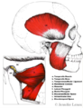
- Ribs (category Chest Wall Anatomy)Louis J. (2001). "Serratus posterior muscles: Anatomy, clinical relevance, and function". Clinical Anatomy. 14 (4): 237–241. doi:10.1002/ca.1039. ISSN 0897-380618 KB (2,468 words) - 20:49, 7 May 2022
- Written by: Dr Jeremy Steinberg; additional contribution by: Dr Amanda Cormack – created: 17 June 2020; last modified: 15 March 2023 This article is still11 KB (1,733 words) - 17:18, 15 March 2023
- threshold of -55mV. Basic Neurophysiology - Members Subpage Spinal Cord Anatomy OpenStax textbook15 KB (1,882 words) - 08:58, 11 April 2023
- judgement and note that this is not necessarily standard practice in NZ. Human anatomy representations typically depict skeletal muscles as separate entities,2 KB (354 words) - 07:00, 22 March 2023
- Hip Labral Tear (section Anatomy)urged to treat the patient not the MRI. Main article: Acetabular Labrum Anatomy The acetabular labrum is a fibrocartilaginous structure that seals the central16 KB (2,524 words) - 07:17, 29 August 2022
- Carpal Tunnel Syndrome (section Anatomy)Ultrasound guided carpal tunnel release is a novel treatment. Knowledge of anatomy variations. Consider the pathophysiology for individual patients to improve20 KB (3,043 words) - 15:17, 11 March 2023
- and "far-out" refers to the site of compression being far lateral. The anatomy of the lumbar spinal canal is divided into the intraspinal canal, the foraminal5 KB (778 words) - 09:59, 17 April 2022
- Hyoid (section Forensic Anatomy of the Hyoid)for a fracture. Part or all of this article or section is derived from Anatomy, Head and Neck, Hyoid Bone by Ghadeer H. AlJulaih; Ritesh G. Menezes., used25 KB (3,213 words) - 20:21, 7 May 2022
- Hip Biomechanics (category Pelvis, Hip, and Thigh Anatomy)This article is still missing information. The hip functions to support the weight of the upper body and transmit forces to the lower extremities. The9 KB (1,251 words) - 16:52, 30 April 2022
- Foot and Ankle Biomechanics (category Foot and Ankle Anatomy)27594929. DOI. Full Text. Ward M Glasoe, H John Yack, Charles L Saltzman, Anatomy and Biomechanics of the First Ray, Physical Therapy, Volume 79, Issue 913 KB (1,816 words) - 08:26, 8 May 2022
- syndrome of LFCN entrapment. Full discussion of injection treatments and the anatomy are discussed elsewhere (See Lateral Femoral Cutaneous Nerve Injection and12 KB (1,419 words) - 09:58, 17 April 2022
- Movements of the Lumbar Spine (category Lumbar Spine Anatomy)in measurement techniques, and researchers not completely appreciating anatomy. In the neutral position the joint runs vertically in the sagittal and coronal15 KB (2,241 words) - 17:34, 30 April 2022
- Ligaments of the Foot and Ankle (category Foot and Ankle Anatomy)This article is still missing information. The ligaments that surround the ankle act to limit plantarflexion and dorsiflexion, anterior and posterior movement9 KB (856 words) - 20:38, 22 March 2023
- This article is still missing information. Foot drop is also known and "drop foot." This is where the patient cannot lift their forefoot secondary to weakness16 KB (2,181 words) - 15:17, 11 March 2023
- Cartoon of pertinent anatomy in femoral nerve block4 KB (493 words) - 19:22, 22 January 2023
- Carpal Instability (section Anatomy)Written by: Dr Jeremy Steinberg – created: 5 August 2021; last modified: 2 March 2023 This page is probably complete! It is awaiting peer review There20 KB (2,468 words) - 16:56, 2 March 2023
- pseudoarthrosis. See images by Chen and colleagues. Transitional Vertebral Anatomy Chen, Zhi-Jia; Cheng, Wei-Jen; Chen, Jean-Lon; Chen, Chien-Hung; Chen, Carl1 KB (131 words) - 16:02, 5 June 2022
- Figure 1: Bone and Ligament Anatomy of the Ankle Joint.18 KB (2,639 words) - 06:00, 2 April 2022
- Gluteal Tendinopathy (section Anatomy)with foot prints of gluteus medius, gluteus minimus, and vastus lateralis Anatomy of the greater trochanter. (a) Three peritrochanteric bursae, (b) osseous29 KB (3,572 words) - 16:24, 23 April 2022
- Shoulder anatomy2 KB (383 words) - 18:45, 5 May 2022
- Anatomy of anterolateral aspect of right knee.2 KB (396 words) - 18:48, 5 May 2022
- Hip anatomy.2 KB (386 words) - 18:47, 5 May 2022
- Management of Lateral Elbow Tendinopathy.pdf Elbow and Forearm Elbow and Forearm Anatomy Elbow and Forearm Conditions Elbow and Forearm Procedures Acute Elbow Pain20 KB (2,909 words) - 10:19, 20 February 2024
- Muscles of the Ankle and Foot (category Foot and Ankle Anatomy)This article is a stub. Strongest plantarflexor - Achilles tendon. Inserts onto the posterior-medial calcaneus. Fires during midstance to slow the forward2 KB (318 words) - 16:56, 30 April 2022
- interlaminar, and caudal injection. A good understanding of lumbar spine anatomy is essential for safe and effective epidural injection. The epidural space30 KB (4,581 words) - 20:40, 22 March 2023
- Cauda Equina Syndrome (section Anatomy and Embryology)Written by: Dr Jeremy Steinberg – created: 15 May 2021; last modified: 11 March 2023 This page is probably complete! It is awaiting peer review Cauda equina21 KB (2,900 words) - 15:21, 11 March 2023
- Shoulder Biomechanics (category Shoulder Anatomy)syndesmosis that doesn't permit much movement. Main article: Glenohumeral Joint Anatomy The GHJ is the synovial ball-and-socket articulation between the humeral22 KB (3,098 words) - 16:58, 30 April 2022
- Thoracic Outlet Syndrome (section Anatomy)individuals. In a study of 50 cadavers only 10% had a bilaterally normal anatomy. Thoracic outlet syndrome is divided into: Neurogenic Thoracic Outlet Syndrome33 KB (4,365 words) - 20:32, 11 March 2023
- Low Back Pain Smoking and Chronic Pain Basic Neurophysiology Spinal Cord Anatomy Hip Osteoarthritis Pompe Disease Tricyclic Antidepressants Neuropathic Pain275 bytes (539 words) - 13:31, 23 April 2022
- tested distal to the dorsal root ganglion due to the unusual cell body anatomy. This discrepancy can be useful in localising the site of a lesion. Motor28 KB (4,121 words) - 16:08, 19 April 2022
- This is based on ported content from Orthopaedia.com It is subject to the CC-BY-NC-SA license. This is an exception to the WikiMSK license. It is not for20 KB (3,049 words) - 20:56, 14 March 2023
- Effective treatment of chronic low back pain in humans reverses abnormal brain anatomy and function. The Journal of neuroscience : the official journal of the20 KB (3,209 words) - 20:47, 22 February 2023
- Nerves of the Lumbar Spine (category Lumbar Spine Anatomy)notes taken from Chapter 10 of: Bogduk, Nikolai. Clinical and radiological anatomy of the lumbar spine. Edinburgh: Elsevier/Churchill Livingstone, 2012. Silverstein28 KB (4,039 words) - 17:34, 30 April 2022
- the sole of the foot, inferior aspect of the toes, and nail beds "Instant Anatomy - Lower Limb - Nerves - Tibial". www.instantanatomy.net. Retrieved 2022-04-172 KB (179 words) - 08:48, 18 April 2022
- unported content Juneja, Pallavi; Munjal, Akul; Hubbard, John B. (2022). "Anatomy, Joints". Treasure Island (FL): StatPearls Publishing. PMID 29939670. Cite1 KB (101 words) - 11:23, 8 May 2022
- unported content Juneja, Pallavi; Munjal, Akul; Hubbard, John B. (2022). "Anatomy, Joints". Treasure Island (FL): StatPearls Publishing. PMID 29939670. Cite1 KB (98 words) - 11:24, 8 May 2022
- images of lunate avascular necrosis. Relate surface anatomy of the wrist to underlying carpal anatomy to facilitate accurate palpation and localization of13 KB (1,703 words) - 19:28, 3 April 2022
- Basic Pulse Sequences for MRI Count vertebra, and assess for transitional anatomy Assess facet joints for effusions, size, cartilage thickness, oedema. Look10 KB (664 words) - 18:36, 20 October 2022
- Lumbar Transforaminal Epidural Steroid Injection/Members
- unported content Juneja, Pallavi; Munjal, Akul; Hubbard, John B. (2022). "Anatomy, Joints". Treasure Island (FL): StatPearls Publishing. PMID 29939670. Cite1 KB (133 words) - 11:23, 8 May 2022
- unported content Juneja, Pallavi; Munjal, Akul; Hubbard, John B. (2022). "Anatomy, Joints". Treasure Island (FL): StatPearls Publishing. PMID 29939670. Cite1 KB (141 words) - 11:23, 8 May 2022
- of maximal tenderness using the Carnett Sign Position: Supine Ultrasound Anatomy Transverse plane in the midline at first to identify the rectus abdominis3 KB (440 words) - 20:09, 27 March 2022
- unported content Juneja, Pallavi; Munjal, Akul; Hubbard, John B. (2022). "Anatomy, Joints". Treasure Island (FL): StatPearls Publishing. PMID 29939670. Cite1 KB (119 words) - 11:23, 8 May 2022
- unported content Juneja, Pallavi; Munjal, Akul; Hubbard, John B. (2022). "Anatomy, Joints". Treasure Island (FL): StatPearls Publishing. PMID 29939670. Cite1 KB (139 words) - 11:23, 8 May 2022
- potential and indications for TFCC repair are influenced by the vascular anatomy of the TFCC. Acute or sub-acute injuries to the peripheral TFCC may be amenable10 KB (1,507 words) - 18:56, 3 April 2022
- of low back pain of primary discal origin” —Cramer G, Darby S. Clinical Anatomy of the Spine, Spinal Cord, and ANS - E-Book. Chapter 7.Elsevier Health Sciences15 KB (2,219 words) - 21:55, 18 March 2022
- 47:137-140. PMID: 28914351. DOI. Bogduk, Nikolai. Clinical and radiological anatomy of the lumbar spine. Chapter 15. Edinburgh: Elsevier/Churchill Livingstone4 KB (654 words) - 20:12, 15 April 2022
- Pelvic anatomy, from theRadiologist5 KB (584 words) - 16:36, 8 May 2021
- exiting L4 nerve root. The relationship between nerve roots and spinal anatomy Less commonly the exiting nerve root is irritated or compressed in the foramen31 KB (4,193 words) - 15:21, 11 March 2023
- obtained. All three radiographs should be examined for a loss of normal anatomy, disruption of the articular surface, involvement of the distal radio-ulnar14 KB (2,070 words) - 06:01, 2 April 2022
- Foot and Ankle Foot and Ankle Anatomy Foot and Ankle Conditions Foot and Ankle Procedures Lower Limb Anatomy Ankle Examination Ankle Pain Differential9 members (4 subcategories, 0 files) - 17:09, 8 May 2021
- values in: |date= (help) Bogduk. Low back pain In: Clinical and Radiological Anatomy of the Lumbar Spine. 5th Edition. Elsevier 2012 Literature Review Reviews2 KB (318 words) - 21:30, 17 April 2022
- cancellous bone. Figure 1: Hindfoot Anatomy Figure 2: Lateral Offset of Calcaneus on Talus Figure 3: Talar Anatomy Figure 4: Os Trigonum (circled in red)19 KB (2,757 words) - 06:02, 2 April 2022
- Hand and Wrist Hand and Wrist Anatomy Hand and Wrist Conditions Hand and Wrist Procedures Hand and Wrist Examination Wrist Pain Differential Diagnoses5 members (3 subcategories, 0 files) - 16:40, 8 May 2021
- Elbow and Forearm Elbow and Forearm Anatomy Elbow and Forearm Conditions Elbow and Forearm Procedures Acute Elbow Pain Elbow Examination Elbow History9 members (3 subcategories, 0 files) - 16:41, 8 May 2021
- Cervical Spine Cervical Spine Anatomy Cervical Spine Conditions Cervical Spine Procedures Acute Neck Pain Anterior Throat Pain Differential Diagnoses Causes11 members (3 subcategories, 0 files) - 19:10, 4 August 2020
- Examination Myotomes Reflex Testing Pain Oriented Sensory Examination Spinal Cord Anatomy Skeletal Muscle Ethan Meltzer. How to Think Like a Neurologist: A Case-Based15 KB (1,556 words) - 20:37, 11 March 2023
- be done in layers. The examiner should be ever-conscious as to the local anatomy. Palpation involves discriminating small differences on a continuum, identifying12 KB (1,617 words) - 13:01, 27 April 2022
- Lumbar Spine Lumbar Spine Anatomy Lumbar Spine Conditions Lumbar Spine Procedures Acute Low Back Pain Causes and Sources of Chronic Low Back Pain Chronic20 members (3 subcategories, 0 files) - 08:55, 14 September 2020
- Category:Torso Anatomy (category Anatomy)1 member (1 subcategory, 0 files) - 16:30, 6 April 2022
- Category:Spine Anatomy (category Anatomy)13 members (7 subcategories, 0 files) - 20:03, 13 July 2020
- Category:Upper Limb Anatomy (category Anatomy)7 members (6 subcategories, 0 files) - 17:02, 30 April 2022
- Category:Lower Limb Anatomy (category Anatomy)7 members (7 subcategories, 0 files) - 16:46, 6 April 2022

File:Anatomy.png (128 × 128 (5 KB)) - 16:55, 3 March 2022- 4 members (0 subcategories, 0 files) - 17:11, 30 April 2022
File:TMJ Anatomy.png File uploaded with MsUpload(518 × 672 (377 KB)) - 09:15, 28 June 2020- 3 members (0 subcategories, 0 files) - 20:49, 7 May 2022
- 7 members (0 subcategories, 0 files) - 17:32, 30 April 2022
- 13 members (0 subcategories, 0 files) - 17:35, 30 April 2022
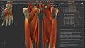
File:Z anatomy screenshot.jpg (1,920 × 1,080 (231 KB)) - 21:11, 6 November 2022
File:Head anatomy drawing.png (225 × 256 (83 KB)) - 08:25, 6 April 2021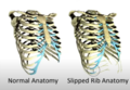
File:Slipped rib anatomy.png File uploaded with MsUpload(607 × 419 (277 KB)) - 06:41, 2 February 2021
File:Cauda equina anatomy.jpg File uploaded with MsUpload(229 × 468 (26 KB)) - 19:57, 16 May 2021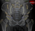
File:Pelvic Xray Anatomy.PNG File uploaded with MsUpload(596 × 527 (521 KB)) - 19:02, 30 August 2020- 3 members (0 subcategories, 0 files) - 17:08, 30 April 2022
- 6 members (0 subcategories, 0 files) - 17:11, 30 April 2022
- 1 member (0 subcategories, 0 files) - 17:08, 30 April 2022
- 9 members (0 subcategories, 0 files) - 17:11, 30 April 2022
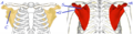
File:Scapula anatomy.png https://orthopaedia.com/page/Scapular-Fractures This work is licensed under a Creative Commons Attribution-NonCommercial-NoDerivatives 4.0 International(624 × 160 (121 KB)) - 20:58, 11 March 2023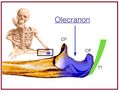
File:Olecranon anatomy.jpg https://orthopaedia.com/page/Olecranon-Fractures This work is licensed under the Creative Commons Attribution-NonCommercial-ShareAlike License.(675 × 518 (43 KB)) - 21:08, 11 March 2023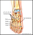
File:Metatarsal anatomy.jpg From https://orthopaedia.com/page/Metatarsal-Fractures This work is licensed under the Creative Commons Attribution-NonCommercial-ShareAlike License.(805 × 847 (82 KB)) - 07:04, 8 March 2022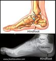
File:Hindfoot anatomy.jpg From https://orthopaedia.com/page/Hindfoot-Fractures This work is licensed under the Creative Commons Attribution-NonCommercial-ShareAlike License.(824 × 900 (88 KB)) - 09:12, 8 March 2022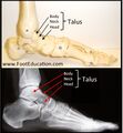
File:Talar anatomy.jpg From https://orthopaedia.com/page/Hindfoot-Fractures This work is licensed under the Creative Commons Attribution-NonCommercial-ShareAlike License.(834 × 900 (89 KB)) - 09:22, 8 March 2022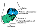
File:Subtalar joint anatomy basic.png File uploaded with MsUpload(724 × 558 (24 KB)) - 13:22, 17 July 2021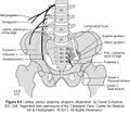
File:Lumbar-Plexus-Anatomy-1.jpg File uploaded with MsUpload(600 × 526 (101 KB)) - 12:36, 16 April 2021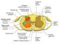
File:Spainal cord sectional anatomy.png File uploaded with MsUpload(1,200 × 880 (124 KB)) - 09:12, 17 May 2021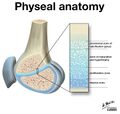
File:Physeal anatomy.jpg Figure 2: Physeal anatomy showing the four zones. (Case courtesy of Dr Matt Skalski, Radiopaedia.org. From the case https://radiopaedia.org/cases/27354)(1,600 × 1,516 (146 KB)) - 19:31, 8 March 2022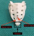
File:Sacral anatomy.jpg From https://resources.wfsahq.org/atotw/ultrasound-guided-caudal-anaesthesia/ This work is licensed under a Creative Commons Attribution-NonCommercial-NoDerivatives(680 × 726 (101 KB)) - 12:18, 30 April 2022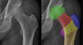
File:Hip xr anatomy.jpg https://orthopaedia.com/page/Hip-Fractures This work is licensed under the Creative Commons Attribution-NonCommercial-ShareAlike License.(1,957 × 1,053 (208 KB)) - 05:16, 13 March 2023- 6 members (3 subcategories, 0 files) - 17:11, 30 April 2022
File:Posterior knee anatomy.png Case courtesy of Assoc Prof Frank Gaillard, Radiopaedia.org. From the case rID: 9330 This work is licensed under the Creative Commons Attribution-NonC(1,200 × 1,200 (172 KB)) - 11:18, 3 August 2021- Thigh Conditions Pelvis, Hip and Thigh Procedures Pelvis, Hip, and Thigh Anatomy Sacroiliac Joint Anterior Thigh Pain Differential Diagnoses Buttock Pain14 members (5 subcategories, 0 files) - 17:19, 8 May 2021
File:Ankle us anatomy.jpg From https://wikem.org/wiki/File:Ankle_us_anatomy.png This work is licensed under the Creative Commons Attribution-ShareAlike 4.0 International License(1,118 × 856 (59 KB)) - 18:35, 5 May 2022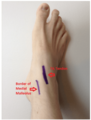
File:Ankle anatomy injection.png From https://wikem.org/wiki/File:Ankle_anatomy_arthrocentesis.png This work is licensed under the Creative Commons Attribution-ShareAlike 4.0 International(726 × 958 (273 KB)) - 19:29, 3 June 2021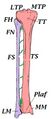
File:Tibia and fibula anatomy.jpg https://orthopaedia.com/page/Tibia-Fractures This work is licensed under the Creative Commons Attribution-NonCommercial-ShareAlike License.(316 × 695 (28 KB)) - 18:33, 13 March 2023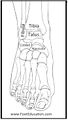
File:Ankle bony anatomy anterior.jpg From https://orthopaedia.com/page/Arthrosis-of-the-Ankle-and-Hindfoot This work is licensed under the Creative Commons Attribution-NonCommercial-ShareAlike(415 × 734 (43 KB)) - 19:53, 8 March 2022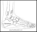
File:Ankle bony anatomy lateral.jpg From https://orthopaedia.com/page/Arthrosis-of-the-Ankle-and-Hindfoot This work is licensed under the Creative Commons Attribution-NonCommercial-ShareAlike(847 × 722 (52 KB)) - 19:53, 8 March 2022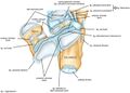
File:Subtalar joint anatomy.jpg Krähenbühl, Nicola et al. “The subtalar joint: A complex mechanism.” EFORT open reviews vol. 2,7 309-316. 6 Jul. 2017, doi:10.1302/2058-5241.2.160050 This(663 × 476 (42 KB)) - 13:17, 17 July 2021
File:S1 TFI anatomy - Tiegs-Heiden 2023.pdf File uploaded with MsUpload(3.86 MB) - 17:21, 15 April 2023
File:Humerus shaft and distal anatomy.jpg https://orthopaedia.com/page/Humeral-Fractures This work is licensed under the Creative Commons Attribution-NonCommercial-ShareAlike License.(1,747 × 869 (113 KB)) - 21:16, 11 March 2023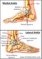
File:Ankle bone and ligament anatomy.jpg From https://orthopaedia.com/page/Ankle-Fractures This work is licensed under the Creative Commons Attribution-NonCommercial-ShareAlike License.(533 × 743 (74 KB)) - 11:13, 8 March 2022
File:Hip anatomy cadaver.jpg From https://commons.wikimedia.org/w/index.php?curid=23768321 This work is licensed under the Creative Commons Attribution-ShareAlike 4.0 International(960 × 720 (96 KB)) - 18:32, 5 May 2022File:Functional Anatomy of the Spine - Bogduk 2016.pdf File uploaded with MsUpload(2.59 MB) - 18:43, 1 September 2021
File:Spinal cord anatomy.jpg Source:Tan S, Faull RLM, Curtis MA. The tracts, cytoarchitecture, and neurochemistry of the spinal cord. Anat Rec (Hoboken). 2023 Apr;306(4):777-819. doi:(1,112 × 1,062 (165 KB)) - 15:56, 10 April 2023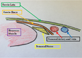
File:Femoral Nerve block anatomy.png From https://wikem.org/wiki/File:Femoral_Nerve_block_anatomy.png This work is licensed under the Creative Commons Attribution-ShareAlike 4.0 International(529 × 376 (323 KB)) - 21:48, 4 May 2022
File:Wrist-anatomy-extrinsic-ligaments.jpg Case courtesy of Dr Matt Skalski, <a href="https://radiopaedia.org/?lang=gb">Radiopaedia.org</a>. From the case <a href="https://radiopaedia.org/cases/43845(1,871 × 1,080 (150 KB)) - 07:18, 6 February 2022File:Surgical anatomy lumbar medial branch neurotomy - Lau 2004.pdf (423 KB) - 19:40, 22 March 2023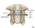
File:Alar-and-cruciform-ligament-anatomy.jpg Case courtesy of Dr Matt Skalski, Radiopaedia.org. From the case rID: 45136 This work is licensed under a Creative Commons Attribution-NonCommercial-NoDerivatives(1,400 × 1,144 (94 KB)) - 09:00, 29 August 2021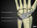
File:Wrist-anatomy-extrinsic-ligaments dorsal.jpg Case courtesy of Dr Matt Skalski, <a href="https://radiopaedia.org/?lang=gb">Radiopaedia.org</a>. From the case <a href="https://radiopaedia.org/cases/43845(1,419 × 1,080 (90 KB)) - 07:22, 6 February 2022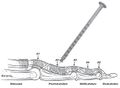
File:Proximal phalanx injection trigger finger anatomy.jpg Kumar et al.. Efficacy of Methylprednisolone Acetate Versus Triamcinolone Acetonide Intra-articular Knee Injection in Patients With Chronic Inflammatory(606 × 446 (45 KB)) - 13:45, 7 June 2021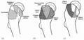
File:Greater trochanter anatomy facets insertions and bursae.jpg Anatomy of the greater trochanter. (a) Three peritrochanteric bursae, (b) osseous facets of the greater trochanter, and (c) insertion sites for the abductor(768 × 369 (48 KB)) - 19:53, 11 April 2022
File:Neck tongue syndrome anatomy.jpg The relationship between the C2 dorsal root ganglion (g), spinal nerve (sn), and ventral ramus (vr, and the lateral atlanto-axial joint (j), posterior(748 × 1,037 (203 KB)) - 10:47, 26 March 2022File:Anatomy and Clinical Importance of the Epidural Space - Fyneface-Ogan 2012.pdf This work is licensed under a Creative Commons Attribution 4.0 International License.(521 KB) - 06:09, 4 January 2022- Category:Muscles of the Torso (category Pelvis, Hip, and Thigh Anatomy)7 members (0 subcategories, 0 files) - 16:29, 6 April 2022
- Category:Muscles of the Lower Limb (category Lower Limb Anatomy)15 members (0 subcategories, 0 files) - 15:59, 15 September 2021
- Figure 1: Bony Anatomy of the Tibia and Fibula. LTP = lateral tibial plateau; MTP = medial tibial plateau; TT = tibial tubercle; TS = tibial shaft; Plaf=19 KB (2,857 words) - 18:41, 13 March 2023
- Category:Nerves of the Lower Limb and Lower Torso (category Lower Limb Anatomy)22 members (0 subcategories, 0 files) - 20:59, 23 April 2022
- Category:Tissues (category Anatomy)13 members (3 subcategories, 0 files) - 17:13, 30 April 2022
- PMCID: PMC6510180. Bogduk. Low back pain In: Clinical and Radiological Anatomy of the Lumbar Spine. 5th Edition. Elsevier 2012 Kapural L, Vrooman B, Sarwar85 KB (12,735 words) - 15:16, 15 April 2023
- treatment is directed more to the soft tissue injury. Main article: Finger Anatomy The phalanges form the fingers and thumb of the hand. The thumb has 2 phalanges10 KB (1,349 words) - 06:04, 2 April 2022
- FRYKHOLM R. Lower cervical vertebrae and intervertebral discs; surgical anatomy and pathology. Acta Chir Scand. 1951;101(5):345-59. PMID: 14868335. Kang11 KB (1,683 words) - 15:30, 11 March 2023
- Category:Biomechanics of the Spine (category Spine Anatomy)3 members (0 subcategories, 0 files) - 17:26, 30 April 2022
- Category:Joints of the Spine (category Spine Anatomy)7 members (0 subcategories, 0 files) - 17:25, 30 April 2022
- Category:Bones of the Spine (category Spine Anatomy)4 members (0 subcategories, 0 files) - 17:25, 30 April 2022
- Category:Muscles of the Spine (category Spine Anatomy)0 members (0 subcategories, 0 files) - 17:26, 30 April 2022
- Category:Nerves of the Spine (category Spine Anatomy)7 members (0 subcategories, 0 files) - 17:26, 30 April 2022
- Category:Regions of the Spine (category Spine Anatomy)2 members (0 subcategories, 0 files) - 17:27, 30 April 2022
- Category:Ligaments of the Spine (category Spine Anatomy)1 member (0 subcategories, 0 files) - 17:33, 30 April 2022
- Category:Regions of the Lower Limb (category Lower Limb Anatomy)3 members (0 subcategories, 0 files) - 16:47, 30 April 2022
- Category:Bones of the Lower Limb (category Lower Limb Anatomy)0 members (0 subcategories, 0 files) - 16:49, 30 April 2022
- Category:Joints of the Lower Limb (category Lower Limb Anatomy)8 members (0 subcategories, 0 files) - 16:49, 30 April 2022
- Category:Joints of the Upper Limb (category Upper Limb Anatomy)9 members (0 subcategories, 0 files) - 17:09, 30 April 2022
- Category:Ligaments of the Lower Limb (category Lower Limb Anatomy)1 member (0 subcategories, 0 files) - 17:18, 30 April 2022
- Category:Bones of the Upper Limb (category Upper Limb Anatomy)0 members (0 subcategories, 0 files) - 17:22, 30 April 2022
- Category:Regions of the Upper Limb (category Upper Limb Anatomy)0 members (0 subcategories, 0 files) - 17:23, 30 April 2022
- Category:Muscles of the Upper Limb (category Upper Limb Anatomy)1 member (0 subcategories, 0 files) - 14:38, 1 August 2022
- Category:Nerves of the Upper Limb (category Upper Limb Anatomy)6 members (0 subcategories, 0 files) - 17:22, 30 April 2022
- Category:Biomechanics of the Lower Limb (category Lower Limb Anatomy)4 members (0 subcategories, 0 files) - 16:52, 30 April 2022
- Category:Biomechanics of the Upper Limb (category Upper Limb Anatomy)3 members (0 subcategories, 0 files) - 17:10, 30 April 2022
- subcategories Knee and Leg Knee and Leg Anatomy Knee and Leg Conditions Knee and Leg Procedures Lower Limb Anatomy Acute Knee Pain Calf Pain Differential4 KB (609 words) - 11:32, 2 April 2022
- https://www.cs.rpi.edu/~kleine/anatomy/bones.html7 members (0 subcategories, 0 files) - 20:48, 7 May 2022
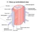
File:Ventrolateral view spinal cord.png Ventrolateral View of the Spinal Cord Anatomy. Source:Tan S, Faull RLM, Curtis MA. The tracts, cytoarchitecture, and neurochemistry of the spinal cord(573 × 496 (153 KB)) - 15:53, 10 April 2023- called endotenon. The surrounding structure is called epitenon. At the gross anatomy level, there is a loose surrounding fascial layer that surrounds some tendons15 KB (2,061 words) - 11:11, 23 March 2023
- By Concept Anatomy and Biomechanics Select [►] to view subcategories Anatomy Joints Lower Limb Anatomy Spine Anatomy Tissues Torso Anatomy Upper Limb Anatomy5 KB (863 words) - 18:15, 12 March 2023
- Knee and Leg Knee and Leg Anatomy Knee and Leg Conditions Knee and Leg Procedures Lower Limb Anatomy Acute Knee Pain Calf Pain Differential Diagnoses Diagnostic13 members (4 subcategories, 0 files) - 15:14, 8 May 2021
- Weakness Myotomes Reflex Testing Pain Oriented Sensory Examination Spinal Cord Anatomy Skeletal Muscle15 KB (1,990 words) - 09:48, 12 March 2023
- Figure 1. Metatarsal Anatomy14 KB (2,086 words) - 06:02, 2 April 2022
- syndrome. All tendon injuries should be examined to determine the specific anatomy that has been disrupted. At the time of initial evaluation, any open injuries14 KB (2,132 words) - 20:19, 3 April 2022
- Shoulder Shoulder Anatomy Shoulder Conditions Shoulder Procedures Shoulder Examination Shoulder Pain Differential Diagnoses5 members (3 subcategories, 0 files) - 19:06, 4 August 2020
- General Spine Lumbar Spine Sacrococcygeal Spine Sacroiliac Joint Spine Anatomy Thoracic Spine7 members (7 subcategories, 0 files) - 19:07, 4 August 2020
- Widespread Anatomy Biomechanics Bone and Cartilage Disorders Communication Examination Functional Approaches Genetic Disorders Lifestyle Neurology Paediatrics22 members (17 subcategories, 0 files) - 19:08, 4 August 2020
- Concepts Anatomy Biomechanics Bone and Cartilage Disorders Clinical Reasoning Communication Evidence Based Medicine Examination Functional Approaches General29 members (29 subcategories, 0 files) - 06:20, 2 April 2022
- Category:Joints (category Anatomy)This category uses the form Joint.26 members (1 subcategory, 0 files) - 20:19, 1 May 2022
- Chest Wall Chest Wall Anatomy Breast Implant Illness Chest Wall Pain Differential Diagnoses First Rib Dysfunction Paediatric Chest Wall Deformities Slipping6 members (1 subcategory, 0 files) - 22:28, 9 April 2022
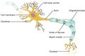
File:The neuron.jpg From OpenStax https://openstax.org/books/anatomy-and-physiology-2e/pages/12-2-nervous-tissue(825 × 552 (50 KB)) - 22:08, 9 August 2023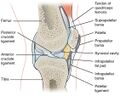
File:Knee Bursae.jpg English: Illustration from Anatomy & Physiology, Connexions Web site. http://cnx.org/content/col11496/1.6/, Jun 19, 2013. This work is licensed under a(1,121 × 899 (116 KB)) - 21:28, 2 August 2021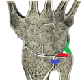
File:TFCC Gray.png Figure 1: Drawing from Gray's Anatomy, showing the normal TFCC in situ: the triangular fibrocartilage disc is shown in red; the ulnocarpal ligament complex(393 × 383 (148 KB)) - 18:55, 3 April 2022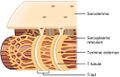
File:T-tubule.jpg From https://openstax.org/books/anatomy-and-physiology/pages/10-2-skeletal-muscle This work is licensed under the Creative Commons Attribution-ShareAlike(577 × 368 (37 KB)) - 15:13, 10 August 2021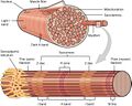
File:Muscle-fibre.jpg From https://openstax.org/books/anatomy-and-physiology/pages/10-2-skeletal-muscle This work is licensed under the Creative Commons Attribution-ShareAlike(801 × 642 (105 KB)) - 15:14, 10 August 2021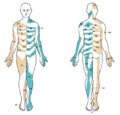
File:Dermatome map lee.PNG From Lee et al.. An evidence-based approach to human dermatomes. Clinical anatomy (New York, N.Y.) 2008. 21:363-73. PMID: 18470936. DOI. The most consistent(677 × 649 (320 KB)) - 06:41, 14 August 2020
File:Motor-end-plate.jpg From https://openstax.org/books/anatomy-and-physiology/pages/10-2-skeletal-muscle This work is licensed under the Creative Commons Attribution-ShareAlike(1,462 × 2,548 (279 KB)) - 15:13, 10 August 2021
File:Sarcomere-binding-sites.jpg From https://openstax.org/books/anatomy-and-physiology/pages/10-2-skeletal-muscle This work is licensed under the Creative Commons Attribution-ShareAlike(830 × 765 (84 KB)) - 15:14, 10 August 2021
File:Endplate fracture consequences.png Own work. Based off Bogduk. Clinical and Radiological Anatomy of the Lumbar Spine. Fifth Edition. 2012 This work is licensed under the Creative Commons(1,920 × 1,309 (114 KB)) - 19:10, 16 September 2021- 27. PMID: 26218010. Bogduk. Low back pain In: Clinical and Radiological Anatomy of the Lumbar Spine. 5th Edition. Elsevier 2012 Manchikanti L, Kosanovic41 KB (6,142 words) - 18:31, 3 November 2022
File:Hierarchical-structure-of-skeletal-muscle.jpg From https://openstax.org/books/anatomy-and-physiology/pages/10-2-skeletal-muscle This work is licensed under the Creative Commons Attribution-ShareAlike(1,435 × 1,482 (220 KB)) - 15:07, 10 August 2021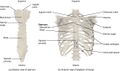
File:Rib Cage.jpg of false ribs are also known as floating ribs (11–12). Illustration from Anatomy & Physiology, Connexions Web site. http://cnx.org/content/col11496/1.6/(1,146 × 678 (284 KB)) - 06:55, 6 June 2021- stopped for 1,500 years. Vesalius broke the dogma of Galenism. He studied anatomy through human dissections (previously disallowed). He published a highly43 KB (6,094 words) - 20:16, 5 December 2022
- epicondyle; (G) olecranon. (Modified from https://www.researchgate.net/figure/Anatomy-of-the-humerus-Modified-from-Wikimedia-This-file-is-licenced-under-the-18 KB (2,454 words) - 08:11, 12 March 2023
File:Finger extensor mechanism dorsal view.jpg Konstantinou P, Pinto I, Karavelis A, Kostretzis L (2017) Extensor Mechanism’s Anatomy at the Metacarpophalangeal Joint. MOJ Orthop Rheumatol 8(4): 00319. DOI:(324 × 532 (23 KB)) - 12:50, 6 February 2022
File:Finger extensor mechanism lateral view.jpg Konstantinou P, Pinto I, Karavelis A, Kostretzis L (2017) Extensor Mechanism’s Anatomy at the Metacarpophalangeal Joint. MOJ Orthop Rheumatol 8(4): 00319. DOI:(558 × 301 (21 KB)) - 12:50, 6 February 2022- Radiculopathy Neuropathic Pain Acute Neck Pain Chronic Neck Pain Bogduk. The anatomy and pathophysiology of neck pain. Physical medicine and rehabilitation clinics41 KB (5,539 words) - 15:21, 11 March 2023
File:GHJ injection under US.jpg Karaca L, Can CE, Pirimoglu B, Tuncer K, Topal M, Okur A, Kantarci M. Anatomy, variants, and pathologies of the superior glenohumeral ligament: magnetic(792 × 418 (41 KB)) - 20:25, 14 February 2022
File:Spinal cord blood supply.jpg Grillo P, Muto M, Garaci F, Muto M, Di Giuliano F. Spinal vascular lesions: anatomy, imaging techniques and treatment. Eur J Radiol Open. 2021 Jul 14;8:100369(1,881 × 1,512 (241 KB)) - 17:33, 4 January 2022- 20 minutes was 0/100, and remained at 0/100 for two hours. Medial branch anatomy AP View The diagnosis is confirmed facetogenic pain arising from the right5 KB (805 words) - 19:00, 22 April 2022
File:TMJ Anatomy2.png File uploaded with MsUpload(622 × 474 (594 KB)) - 09:18, 28 June 2020
File:TMJ Anatomy3.png File uploaded with MsUpload(612 × 547 (177 KB)) - 09:23, 28 June 2020- iliac spine, by the pull of the rectus femoris. (Annotation of a Gray's Anatomy original)23 KB (2,933 words) - 08:10, 12 March 2023
- formation. Because the bone scan assesses a metabolic response (and not static anatomy), positive findings may not be apparent for up to 72 hours after the administration28 KB (3,720 words) - 05:24, 13 March 2023
- Brismée J-M, Sizer PS, Courtney CA. Temporomandibular disorders. Part 1: anatomy and examination/diagnosis. J Man Manip Ther. 2014 Feb;22(1):2–12. Ren YF35 KB (5,034 words) - 19:43, 6 January 2023


